

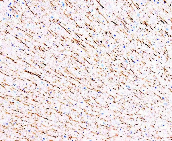
IHC-P analysis of rat brain tissue using GTX11264 MAP1A antibody [HM-1].
IHC-P analysis of rat brain tissue using GTX11264 MAP1A antibody [HM-1].
MAP1A antibody [HM-1]
GTX11264
ApplicationsImmunoPrecipitation, Western Blot, ImmunoHistoChemistry, ImmunoHistoChemistry Frozen, ImmunoHistoChemistry Paraffin
Product group Antibodies
ReactivityMouse, Rat
TargetMap1a
Overview
- SupplierGeneTex
- Product NameMAP1A antibody [HM-1]
- Delivery Days Customer9
- Application Supplier NoteWB: 0.5-2microg/ml. IHC-P: 1-2microg/ml. IHC-Fr: 1-2microg/ml. *Optimal dilutions/concentrations should be determined by the researcher.Not tested in other applications.
- ApplicationsImmunoPrecipitation, Western Blot, ImmunoHistoChemistry, ImmunoHistoChemistry Frozen, ImmunoHistoChemistry Paraffin
- CertificationResearch Use Only
- ClonalityMonoclonal
- Clone IDHM-1
- Concentration0.1 mg/ml
- ConjugateUnconjugated
- Gene ID25152
- Target nameMap1a
- Target descriptionmicrotubule-associated protein 1A
- Target synonymsMtap1a, microtubule-associated protein 1A, MAP-1A
- HostMouse
- IsotypeIgG1
- Protein IDP34926
- Protein NameMicrotubule-associated protein 1A
- Scientific DescriptionMicrotubules are the ubiquitous cytoskeletal structural components that are involved in intracellular transport. They are composed of tubulin and microtubule-associated proteins (MAPs). There is considerable evidence that MAPs may mediate the binding of membranous organelles, actin filaments and intermediate filaments to microtubules, leading to the speculation that they may therefore be important for cellular processes such as mitosis and organelle transport, and for determining the dynamic properties of the cytoskeleton. Two classes of high molecular weight components termed MAP1 and MAP2 have been demonstrated to co-purify with tubulin during cycles of microtubule assembly and disassembly, and to stimulate microtubule assembly in vitro. MAP1 is one of the major neuronal MAPs as well as being the largest (350 kD). Purified preparations of MAP1 from bovine brain have been demonstrated to contain at least two low molecualr weight components (19-34 kD) that remain tightly associated with MAP1 heavy chains under nondenaturing conditions. In contrast to MAP2 which is localized primarily in the dendrites of neurons in brain and possibly in small amounts in other cells, MAP1 is more generally distributed, being found in both dendrites and axons of neurons and in glial cells in brain, in chromtophores, and on both interphase and mitotic microtubules in various tissue culture cells, suggesting that MAP1 may have a more general function. In the newborn rat brain the expression of MAP1 is almost absent. Its levels begin to increase from postnatal day 5 and increase steadily, in step with neuronal differentiation, reaching a maximum around postnatal day 28, the time when neurons have reached their mature morphology. MAP1 is degraded by a Cathepsin D like protease in the brain of aged rats. In developmental neurobiology MAP1 acts as a marker of neuronal maturation.
- ReactivityMouse, Rat
- Storage Instruction-20°C or -80°C,2°C to 8°C
- UNSPSC12352203
References
- Murakami N, Bolton DC, Kida E, et al. Phosphorylation by Dyrk1A of clathrin coated vesicle-associated proteins: identification of the substrate proteins and the effects of phosphorylation. PLoS One. 2012,7(4):e34845. doi: 10.1371/journal.pone.0034845Read this paper
Datasheet
Related products
Product group Antibodies
ApplicationsImmunoPrecipitation, Western Blot, ImmunoCytoChemistry, ImmunoHistoChemistry
ReactivityRat
TargetMap1a
- SizePrice