

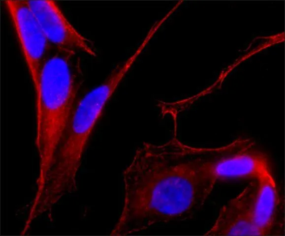
ICC/IF analysis of SH-SY-5Y cells using GTX11268 MAP2 antibody [AP-20] at 1:500(red) with DAPI(blue).Cells were fixed and permeabilized with methanol followed by methanol:acetone.
![ICC/IF analysis of B35 cells using GTX11268 MAP2 antibody [AP-20] at 1:100(red) with DAPI(blue).Cells were fixed and permeabilized with methanol followed by methanol:acetone.](https://www.genetex.com/upload/website/prouct_img/normal/GTX11268/GTX11268_20170605_ICCIF_w_23060500_350.webp "ICC/IF analysis of B35 cells using GTX11268 MAP2 antibody [AP-20] at 1:100(red) with DAPI(blue).Cells were fixed and permeabilized with methanol followed by methanol:acetone.")
![WB analysis of rat brain extract using GTX11268 MAP2 antibody[AP-20]. Lane A : Antibody dilution 1:500 Lane B : Secondary antibody only](https://www.genetex.com/upload/website/prouct_img/normal/GTX11268/GTX11268_20170605_WB_w_23060500_526.webp "WB analysis of rat brain extract using GTX11268 MAP2 antibody[AP-20]. Lane A : Antibody dilution 1:500 Lane B : Secondary antibody only")
ICC/IF analysis of SH-SY-5Y cells using GTX11268 MAP2 antibody [AP-20] at 1:500(red) with DAPI(blue).Cells were fixed and permeabilized with methanol followed by methanol:acetone.
MAP2 antibody [AP-20]
GTX11268
ApplicationsImmunoFluorescence, Western Blot, ImmunoCytoChemistry, ImmunoHistoChemistry, ImmunoHistoChemistry Paraffin
Product group Antibodies
ReactivityAmphibian, Avian, Bovine, Human, Mouse, Rat, Xenopus
TargetMAP2
Overview
- SupplierGeneTex
- Product NameMAP2 antibody [AP-20]
- Delivery Days Customer9
- Application Supplier NoteWB: 1:500. *Optimal dilutions/concentrations should be determined by the researcher.Not tested in other applications.
- ApplicationsImmunoFluorescence, Western Blot, ImmunoCytoChemistry, ImmunoHistoChemistry, ImmunoHistoChemistry Paraffin
- CertificationResearch Use Only
- ClonalityMonoclonal
- Clone IDAP-20
- ConjugateUnconjugated
- Gene ID281294
- Target nameMAP2
- Target descriptionmicrotubule associated protein 2
- Target synonymsmicrotubule-associated protein 2, microtubule associated protein 2C
- HostMouse
- IsotypeIgG1
- Scientific DescriptionMAP2 is the major microtubule associated protein of brain tissue. There are three forms of MAP2; two are similarily sized with apparent molecular weights of 280 kDa (MAP2a and MAP2b) and the third with a lower molecular weight of 70 kDa (MAP2c). In the newborn rat brain, MAP2b and MAP2c are present, while MAP2a is absent. Between postnatal days 10 and 20, MAP2a appears. At the same time, the level of MAP2c drops by 10-fold. This change happens during the period when dendrite growth is completed and when neurons have reached their mature morphology. MAP2 is degraded by a Cathepsin D-like protease in the brain of aged rats. There is some indication that MAP2 is expressed at higher levels in some types of neurons than in other types. MAP2 is known to promote microtubule assembly and to form side-arms on microtubules. It also interacts with neurofilaments, actin, and other elements of the cytoskeleton.
- ReactivityAmphibian, Avian, Bovine, Human, Mouse, Rat, Xenopus
- Storage Instruction-20°C or -80°C,2°C to 8°C
- UNSPSC12352203
References
- Valle-Bautista R, Márquez-Valadez B, Fragoso-Cabrera AD, et al. Impaired Cortical Cytoarchitecture and Reduced Excitability of Deep-Layer Neurons in the Offspring of Diabetic Rats. Front Cell Dev Biol. 2020,8:564561. doi: 10.3389/fcell.2020.564561Read this paper
- Tian E, Sun G, et al. Small-Molecule-Based Lineage Reprogramming Creates Functional Astrocytes. Cell Rep. 2016,16(3):781-92. doi: 10.1016/j.celrep.2016.06.042Read this paper
Datasheet
Related products
Product group Antibodies
MAP2 antibody [MT-08]GTX79911
ApplicationsImmunoFluorescence, ImmunoPrecipitation, Western Blot, ELISA, ImmunoCytoChemistry, ImmunoHistoChemistry, ImmunoHistoChemistry Frozen, ImmunoHistoChemistry Paraffin
ReactivityBovine, Human, Mouse, Porcine
TargetMAP2
- SizePrice
Product group Antibodies
MAP2 antibody [MT-07]GTX79912
ApplicationsImmunoFluorescence, ImmunoPrecipitation, Western Blot, ELISA, ImmunoCytoChemistry, ImmunoHistoChemistry, ImmunoHistoChemistry Frozen, ImmunoHistoChemistry Paraffin
ReactivityBovine, Human, Mouse, Porcine
TargetMAP2
- SizePrice
![WB analysis of microtubules partially purified from pig brain lysate using GTX27756 MAP2 antibody [MT-01]. Lane 1 : GTX27756 Lane 2 : GTX79912 Lane 3 : GTX79911](https://www.genetex.com/upload/website/prouct_img/normal/GTX27756/GTX27756_20191025_AP_001_193_w_23060722_809.webp)
Product group Antibodies
References
MAP2 antibody [MT-01]GTX27756
ApplicationsImmunoFluorescence, ImmunoPrecipitation, Western Blot, ImmunoCytoChemistry
ReactivityBovine, Feline, Human, Mouse, Porcine, Rat
TargetMAP2
- SizePrice