


Products
Are you looking for life science and diagnostic reagents? We offer one of the most extensive ranges in the Benelux. There are currently more than 8 million products in our webshop, which are manufactured by more than 130 suppliers. We hope to support your research with everything you need.

Product group Antibodies
Anti-GPBAR AntibodyA30825
ApplicationsWestern Blot, ELISA, ImmunoHistoChemistry
TargetGPBAR1
- SizePrice

Product group Antibodies
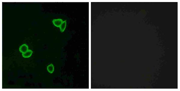
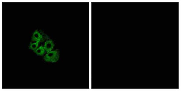
Anti-GPRC5B AntibodyA30826
ApplicationsImmunoFluorescence, Western Blot, ELISA, ImmunoCytoChemistry
TargetGPRC5B
- SizePrice

Product group Antibodies
Anti-GPRC5C AntibodyA30827
ApplicationsImmunoFluorescence, Western Blot, ELISA, ImmunoCytoChemistry
TargetGPRC5C
- SizePrice

Product group Antibodies
Anti-GPRC5D AntibodyA30828
ApplicationsImmunoFluorescence, Western Blot, ELISA, ImmunoCytoChemistry
TargetGPRC5D
- SizePrice

Product group Antibodies
Anti-GPRC6A AntibodyA30829
ApplicationsImmunoFluorescence, Western Blot, ELISA, ImmunoCytoChemistry
TargetGPRC6A
- SizePrice

Product group Antibodies
ApplicationsImmunoFluorescence, Western Blot, ELISA, ImmunoCytoChemistry
TargetADGRG1
- SizePrice

Product group Antibodies
Anti-GPR92 AntibodyA30831
ApplicationsImmunoFluorescence, Western Blot, ELISA, ImmunoCytoChemistry, ImmunoHistoChemistry
TargetLPAR5
- SizePrice
Product group Antibodies
ApplicationsWestern Blot, ELISA, ImmunoHistoChemistry
TargetADGRL1
- SizePrice

Product group Antibodies
ApplicationsImmunoFluorescence, ELISA, ImmunoCytoChemistry, ImmunoHistoChemistry
TargetADGRL2
- SizePrice
Product group Antibodies
Anti-LSHR AntibodyA30834
ApplicationsWestern Blot, ELISA, ImmunoHistoChemistry
TargetLHCGR
- SizePrice

Product group Antibodies
Anti-MPRA AntibodyA30835
ApplicationsWestern Blot, ImmunoHistoChemistry
TargetPAQR7
- SizePrice

Product group Antibodies
Anti-MPRG PAQR5 AntibodyA30836
ApplicationsWestern Blot, ELISA, ImmunoHistoChemistry
TargetPAQR5
- SizePrice
Didn't find what you were looking for?
Search through our product groups to find the right product
Back to overview