


Products
Are you looking for life science and diagnostic reagents? We offer one of the most extensive ranges in the Benelux. There are currently more than 8 million products in our webshop, which are manufactured by more than 130 suppliers. We hope to support your research with everything you need.
Product group Antibodies
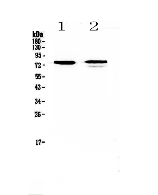
Anti-FZD3 Antibody Picoband(r)A04680-1-HRP
ApplicationsWestern Blot, ImmunoHistoChemistry
ReactivityHuman, Mouse, Rat
TargetFZD3
- SizePrice
Product group Antibodies
Anti-FZD3 Antibody Picoband(r)A04680-1-IFLUOR647
ApplicationsWestern Blot, ImmunoHistoChemistry
ReactivityHuman, Mouse, Rat
TargetFZD3
- SizePrice
Product group Antibodies
Anti-FZD3 Antibody Picoband(r)A04680-1-PE
ApplicationsWestern Blot, ImmunoHistoChemistry
ReactivityHuman, Mouse, Rat
TargetFZD3
- SizePrice
Product group Antibodies
Anti-FZD3 Antibody Picoband(r)A04680-1
ApplicationsWestern Blot, ImmunoHistoChemistry
ReactivityHuman, Mouse, Rat
TargetFZD3
- SizePrice
Product group Antibodies
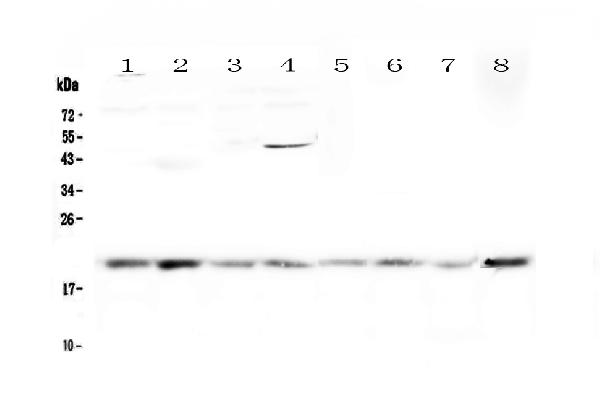
Anti-GADD45G Antibody Picoband(r)A04681-1-10UG
ApplicationsWestern Blot
ReactivityHuman, Mouse, Rat
TargetGADD45G
- SizePrice
Product group Antibodies
Anti-GADD45G Antibody Picoband(r)A04681-1-APC
ApplicationsWestern Blot
ReactivityHuman, Mouse, Rat
TargetGADD45G
- SizePrice
Product group Antibodies
Anti-GADD45G Antibody Picoband(r)A04681-1-BIOTIN
ApplicationsWestern Blot
ReactivityHuman, Mouse, Rat
TargetGADD45G
- SizePrice
Product group Antibodies
Anti-GADD45G Antibody Picoband(r)A04681-1-CARRIER-FREE
ApplicationsWestern Blot
ReactivityHuman, Mouse, Rat
TargetGADD45G
- SizePrice
Product group Antibodies
Anti-GADD45G Antibody Picoband(r)A04681-1-CY3
ApplicationsWestern Blot
ReactivityHuman, Mouse, Rat
TargetGADD45G
- SizePrice
Product group Antibodies
Anti-GADD45G Antibody Picoband(r)A04681-1-DYLIGHT488
ApplicationsWestern Blot
ReactivityHuman, Mouse, Rat
TargetGADD45G
- SizePrice
Product group Antibodies
Anti-GADD45G Antibody Picoband(r)A04681-1-DYLIGHT550
ApplicationsWestern Blot
ReactivityHuman, Mouse, Rat
TargetGADD45G
- SizePrice
Product group Antibodies
Anti-GADD45G Antibody Picoband(r)A04681-1-DYLIGHT594
ApplicationsWestern Blot
ReactivityHuman, Mouse, Rat
TargetGADD45G
- SizePrice
Didn't find what you were looking for?
Search through our product groups to find the right product
Back to overview