


Products
Are you looking for life science and diagnostic reagents? We offer one of the most extensive ranges in the Benelux. There are currently more than 8 million products in our webshop, which are manufactured by more than 130 suppliers. We hope to support your research with everything you need.

Product group Antibodies
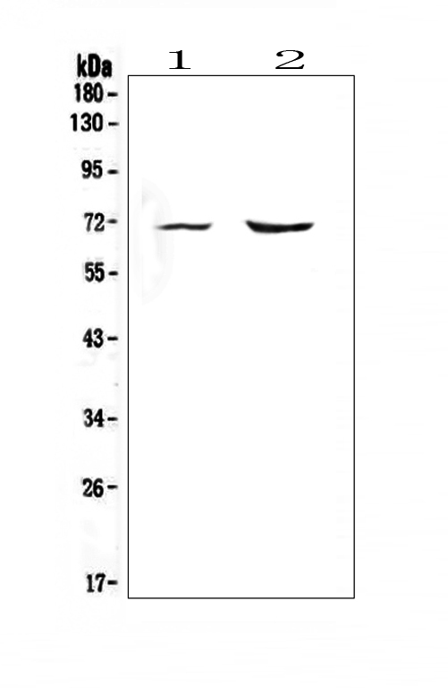
Anti-KCNQ2 Antibody Picoband(r)A01259-1-FITC
ApplicationsFlow Cytometry, Western Blot, ELISA, ImmunoHistoChemistry
ReactivityHuman, Mouse, Rat
TargetKCNQ2
- SizePrice
Product group Antibodies
Anti-KCNQ2 Antibody Picoband(r)A01259-1-HRP
ApplicationsFlow Cytometry, Western Blot, ELISA, ImmunoHistoChemistry
ReactivityHuman, Mouse, Rat
TargetKCNQ2
- SizePrice
Product group Antibodies
Anti-KCNQ2 Antibody Picoband(r)A01259-1-IFLUOR647
ApplicationsFlow Cytometry, Western Blot, ELISA, ImmunoHistoChemistry
ReactivityHuman, Mouse, Rat
TargetKCNQ2
- SizePrice
Product group Antibodies
Anti-KCNQ2 Antibody Picoband(r)A01259-1-PE
ApplicationsFlow Cytometry, Western Blot, ELISA, ImmunoHistoChemistry
ReactivityHuman, Mouse, Rat
TargetKCNQ2
- SizePrice
Product group Antibodies
Anti-KCNQ2 Antibody Picoband(r)A01259-1
ApplicationsFlow Cytometry, Western Blot, ELISA, ImmunoHistoChemistry
ReactivityHuman, Mouse, Rat
TargetKCNQ2
- SizePrice
Product group Antibodies
ApplicationsImmunoFluorescence, ELISA, ImmunoHistoChemistry
ReactivityHuman, Mouse, Rat
TargetKCNQ2
- SizePrice
Product group Antibodies
ApplicationsImmunoFluorescence, Western Blot, ELISA, ImmunoHistoChemistry
ReactivityHuman, Mouse, Rat
TargetKCNQ2
- SizePrice
Product group Antibodies
ApplicationsWestern Blot, ELISA
ReactivityHuman, Mouse
TargetCFB
- SizePrice

Product group Antibodies
Anti-SDHD AntibodyA01261
ApplicationsImmunoFluorescence, Western Blot, ELISA, ImmunoCytoChemistry
ReactivityBovine, Human, Mouse, Porcine, Rat, Sheep
TargetSDHD
- SizePrice
Product group Antibodies
Anti-Melanoma gp100/PMEL Antibody Picoband(r)A01262-1-10UG
ApplicationsWestern Blot
ReactivityHuman, Mouse, Rat
TargetPMEL
- SizePrice
Product group Antibodies
Anti-Melanoma gp100/PMEL Antibody Picoband(r)A01262-1-APC
ApplicationsWestern Blot
ReactivityHuman, Mouse, Rat
TargetPMEL
- SizePrice
Product group Antibodies
Anti-Melanoma gp100/PMEL Antibody Picoband(r)A01262-1-BIOTIN
ApplicationsWestern Blot
ReactivityHuman, Mouse, Rat
TargetPMEL
- SizePrice
Didn't find what you were looking for?
Search through our product groups to find the right product
Back to overview