


Products
Are you looking for life science and diagnostic reagents? We offer one of the most extensive ranges in the Benelux. There are currently more than 8 million products in our webshop, which are manufactured by more than 130 suppliers. We hope to support your research with everything you need.

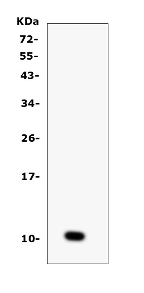
Product group Antibodies
References
ApplicationsWestern Blot, ImmunoHistoChemistry
ReactivityHuman, Virus
TargetS
- SizePrice
Product group Antibodies
Anti-CCL6 Antibody Picoband(r)A30384-10UG
ApplicationsWestern Blot, ELISA, ImmunoHistoChemistry
ReactivityMouse, Rat
TargetCcl6
- SizePrice
Product group Antibodies
Anti-CCL6 Antibody Picoband(r)A30384-APC
ApplicationsWestern Blot, ELISA, ImmunoHistoChemistry
ReactivityMouse, Rat
TargetCcl6
- SizePrice
Product group Antibodies
Anti-CCL6 Antibody Picoband(r)A30384-BIOTIN
ApplicationsWestern Blot, ELISA, ImmunoHistoChemistry
ReactivityMouse, Rat
TargetCcl6
- SizePrice
Product group Antibodies
Anti-CCL6 Antibody Picoband(r)A30384-CARRIER-FREE
ApplicationsWestern Blot, ELISA, ImmunoHistoChemistry
ReactivityMouse, Rat
TargetCcl6
- SizePrice
Product group Antibodies
Anti-CCL6 Antibody Picoband(r)A30384-CY3
ApplicationsWestern Blot, ELISA, ImmunoHistoChemistry
ReactivityMouse, Rat
TargetCcl6
- SizePrice
Product group Antibodies
Anti-CCL6 Antibody Picoband(r)A30384-DYLIGHT488
ApplicationsWestern Blot, ELISA, ImmunoHistoChemistry
ReactivityMouse, Rat
TargetCcl6
- SizePrice
Product group Antibodies
Anti-CCL6 Antibody Picoband(r)A30384-DYLIGHT550
ApplicationsWestern Blot, ELISA, ImmunoHistoChemistry
ReactivityMouse, Rat
TargetCcl6
- SizePrice
Product group Antibodies
Anti-CCL6 Antibody Picoband(r)A30384-DYLIGHT594
ApplicationsWestern Blot, ELISA, ImmunoHistoChemistry
ReactivityMouse, Rat
TargetCcl6
- SizePrice
Product group Antibodies
Anti-CCL6 Antibody Picoband(r)A30384-FITC
ApplicationsWestern Blot, ELISA, ImmunoHistoChemistry
ReactivityMouse, Rat
TargetCcl6
- SizePrice
Product group Antibodies
Anti-CCL6 Antibody Picoband(r)A30384-HRP
ApplicationsWestern Blot, ELISA, ImmunoHistoChemistry
ReactivityMouse, Rat
TargetCcl6
- SizePrice
Product group Antibodies
Anti-CCL6 Antibody Picoband(r)A30384-IFLUOR647
ApplicationsWestern Blot, ELISA, ImmunoHistoChemistry
ReactivityMouse, Rat
TargetCcl6
- SizePrice
Didn't find what you were looking for?
Search through our product groups to find the right product
Back to overview