Bio-Connect
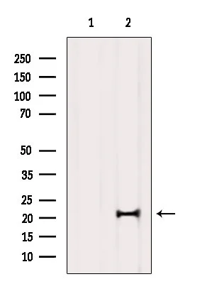
WB analysis of mouse brain tissue lysate using GTX00806 MED29 antibody. The lane on the left was treated with blocking peptide.

MED29 antibody

GTX00806
GeneTex
ApplicationsWestern Blot
Product group Antibodies
TargetMED29
Sign in to order and to see your custom pricing.
Large volume orders?
Order with a bulk request

Overview

  • Supplier
    GeneTex
  • Product Name
    MED29 antibody
  • Delivery Days Customer
    9
  • Application Supplier Note
    WB: 1:1000-1:3000. *Optimal dilutions/concentrations should be determined by the researcher.Not tested in other applications.
  • Applications
    Western Blot
  • Certification
    Research Use Only
  • Clonality
    Polyclonal
  • Concentration
    1 mg/ml
  • Conjugate
    Unconjugated
  • Gene ID55588
  • Target name
    MED29
  • Target description
    mediator complex subunit 29
  • Target synonyms
    intersex-like protein; IXL; MED2; mediator of RNA polymerase II transcription subunit 29
  • Host
    Rabbit
  • Isotype
    IgG
  • Protein IDQ9NX70
  • Protein Name
    Mediator of RNA polymerase II transcription subunit 29
  • Scientific Description
    MED29 is a subunit of the Mediator complex, a multiprotein coactivator of RNA transcription that interacts with DNA-bound transcriptional activators, RNA polymerase II (see MIM 180660), and general initiation factors (Sato et al., 2003 [PubMed 14576168]).[supplied by OMIM, Aug 2009]
  • Storage Instruction
    -20°C or -80°C,2°C to 8°C
  • UNSPSC
    12352203

Related products

Anti-MED29 Antibody
Product group Antibodies
Antibodies.com
ApplicationsWestern Blot
  • SizePrice
Figure 1. Western blot analysis of MED29 using anti-MED29 antibody (A10590-1). Electrophoresis was performed on a 5-20% SDS-PAGE gel at 70V (Stacking gel) / 90V (Resolving gel) for 2-3 hours. The sample well of each lane was loaded with 30 ug of sample under reducing conditions. Lane 1: human Siha whole cell lysates, Lane 2: human RT4 whole cell lysates, Lane 3: human MCF-7 whole cell lysates, Lane 4: human 293T whole cell lysates, Lane 5: human Jurkat whole cell lysates, Lane 6: human U251 whole cell lysates, Lane 7: human Caco-2 whole cell lysates. After electrophoresis, proteins were transferred to a nitrocellulose membrane at 150 mA for 50-90 minutes. Blocked the membrane with 5% non-fat milk/TBS for 1.5 hour at RT. The membrane was incubated with rabbit anti-MED29 antigen affinity purified polyclonal antibody (Catalog # A10590-1) at 0.5 microg/mL overnight at 4°C, then washed with TBS-0.1%Tween 3 times with 5 minutes each and probed with a goat anti-rabbit IgG-HRP secondary antibody at a dilution of 1:5000 for 1.5 hour at RT. The signal is developed using an Enhanced Chemiluminescent detection (ECL) kit (Catalog # EK1002) with Tanon 5200 system. A specific band was detected for MED29 at approximately 21 kDa. The expected band size for MED29 is at 21 kDa.
Product group Antibodies
Boster Bio
Anti-MED29 Antibody Picoband(r)A10590-1-CARRIER-FREE
ApplicationsImmunoFluorescence, Western Blot, ELISA, ImmunoCytoChemistry
TargetMED29
  • SizePrice
Immunofluorescent staining of human cell line A-431 shows localization to nucleoplasm.
Product group Antibodies
Atlas Antibodies
ApplicationsImmunoCytoChemistry
TargetMED29
  • SizePrice
Product group Antibodies
LifeSpan BioSciences
ApplicationsWestern Blot
TargetMED29
  • SizePrice
Product group Antibodies
Cusabio
MED29 AntibodyCSB-PA885752XA01HU
ApplicationsWestern Blot, ELISA
ReactivityHuman
TargetMED29
  • SizePrice