

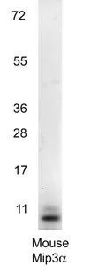
Western blot using GeneTex's anti-Mouse MIP3a antibody shows detection of a band ~11 kDa in size corresponding to recombinant mouse MIP3a. Recombinant mouse MIP-3a was loaded on to an SDS-PAGE gel at 0.25 μg and after separation was transferred to nitrocellulose. The membrane was blocked with 1% BSA in TBST for 30 min at RT, followed by incubation with primary antibody diluted 1:1,000 in 1% BSA in TBST overnight at 4oC. After washes, the blot was reacted with secondary antibody HRP Goat anti-Rabbit IgG antibody diluted 1:40,000 in blocking buffer) for 30 min at RT followed by reation with FemtoMax Chemiluminescent substrate. Data was collected using Bio-Rad VersaDocR 4000 MP imaging system.

Western blot using GeneTex's anti-Mouse MIP3a antibody shows detection of a band ~11 kDa in size corresponding to recombinant mouse MIP3a. Recombinant mouse MIP-3a was loaded on to an SDS-PAGE gel at 0.25 μg and after separation was transferred to nitrocellulose. The membrane was blocked with 1% BSA in TBST for 30 min at RT, followed by incubation with primary antibody diluted 1:1,000 in 1% BSA in TBST overnight at 4oC. After washes, the blot was reacted with secondary antibody HRP Goat anti-Rabbit IgG antibody diluted 1:40,000 in blocking buffer) for 30 min at RT followed by reation with FemtoMax Chemiluminescent substrate. Data was collected using Bio-Rad VersaDocR 4000 MP imaging system.
MIP3 alpha antibody
GTX48690
ApplicationsWestern Blot, ELISA
Product group Antibodies
ReactivityMouse, Rat
TargetCcl20
Overview
- SupplierGeneTex
- Product NameMIP3 alpha antibody
- Delivery Days Customer9
- Application Supplier NoteWB: 1:1000. ELISA: 1:10000. *Optimal dilutions/concentrations should be determined by the researcher.Not tested in other applications.
- ApplicationsWestern Blot, ELISA
- CertificationResearch Use Only
- ClonalityPolyclonal
- Concentration1 mg/ml
- ConjugateUnconjugated
- Gene ID20297
- Target nameCcl20
- Target descriptionC-C motif chemokine ligand 20
- Target synonymsCKb4, LARC, MIP-3A, MIP-3[a], MIP3A, ST38, Scya20, exodus-1, C-C motif chemokine 20, CC chemokine LARC, CC chemokine ST38, MIP-3-alpha, beta-chemokine exodus-1, chemokine (C-C motif) ligand 20, liver and activation-regulated chemokine, macrophage inflammatory protein 3 alpha, small inducible cytokine subfamily A20, small-inducible cytokine A20
- HostRabbit
- IsotypeIgG
- Protein IDO89093
- Protein NameC-C motif chemokine 20
- Scientific DescriptionMIP-3a (also known as C-C motif chemokine 20, small-inducible cytokine A20, macrophage inflammatory protein 3 alpha, MIP-3-alpha, liver and activation-regulated chemokine, CC chemokine LARC and beta chemokine exodus-1) is a chemotactic factor that attracts lymphocytes and, slightly, neutrophils, but not monocytes. MIP-3a inhibits proliferation of myeloid progenitors in colony formation assays and may be involved in formation and function of the mucosal lymphoid tissues by attracting lymphocytes and dendritic cells towards epithelial cells. C-terminal processed forms have been shown to be equally chemotactically active for leukocytes. MIP-3a also possesses antibacterial activity against E.coli and S.aureus. MIP-3a is a secreted protein that is expressed predominantly in the liver, lymph nodes, appendix, peripheral blood lymphocytes, and fetal lung. Low levels of expression are also seen in thymus, prostate, testis, small intestine and colon. C-terminal processed forms which lack 1, 3 or 6 amino acids are produced by proteolytic cleavage after secretion from peripheral blood monocytes.
- ReactivityMouse, Rat
- Storage Instruction-20°C or -80°C,2°C to 8°C
- UNSPSC41116161
Datasheet
Related products

Product group Antibodies
Anti-MIP-3 Alpha/CCL20 Antibody Picoband(r)A00748-2-CARRIER-FREE
ApplicationsWestern Blot, ELISA
ReactivityMouse, Rat
TargetCcl20
- SizePrice
Product group Antibodies

Anti-CCL20 [AB1]AB03417-1.1-BT
ApplicationsELISA, Other Application
ReactivityMouse
TargetCcl20
- SizePrice
Product group Antibodies
ApplicationsImmunoPrecipitation, Western Blot, ImmunoCytoChemistry, ImmunoHistoChemistry
ReactivityMouse
TargetCcl20
- SizePrice
Product group Antibodies
MIP3 alpha antibody (Biotin)GTX48688
ApplicationsWestern Blot, ELISA, ImmunoHistoChemistry
ReactivityMouse, Rat
TargetCcl20
- SizePrice
Product group Antibodies
MIP3 alpha antibody (HRP)GTX48689
ApplicationsWestern Blot, ELISA, ImmunoHistoChemistry
ReactivityMouse, Rat
TargetCcl20
- SizePrice
Product group Antibodies
MIP3 alpha antibody [11J22]GTX53264
ApplicationsWestern Blot, Neutralisation/Blocking
ReactivityMouse
TargetCcl20
- SizePrice
Product group Antibodies
References
CCL20 Polyclonal AntibodyBS-1268R
ApplicationsImmunoFluorescence, ELISA, ImmunoCytoChemistry, ImmunoHistoChemistry, ImmunoHistoChemistry Frozen, ImmunoHistoChemistry Paraffin
ReactivityHuman, Mouse, Rat
TargetCcl20
- SizePrice