

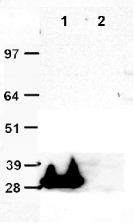
Immunoprecipitation (IP) was performed with overexpression lysate expressed with tGFP expression vector (Cat# PS100010) or control lysate transfected with pCMV-Entry empty vector (Cat# PS100001). Briefly, 20uL anti-tGFP (2H8) magnetic beads (Cat# TA150039) were incubated with tGFP overexpression lysate or negative control lysate overnight at 4°C, respectively. After washing three times with RIPA buffer, 20uL 4x SDS sample buffer was applied to the lysate-beads mixture. The samples were boiled at 95°C for 10min. The IP sample supernatants were fractionated on SDS-PAGE gel, blotted onto nitrocellulose membrane. The IP efficiency was evaluated by using HRP-conjugated anti-tGFP antibody (Cat# TA150043). Lane 1: anti-tGFP (2H8) magnetic beads (20uL) with tGFP expression lysate; Lane 2: anti-tGFP magnetic beads (20uL) with negative control lysate.
Immunoprecipitation (IP) was performed with overexpression lysate expressed with tGFP expression vector (Cat# PS100010) or control lysate transfected with pCMV-Entry empty vector (Cat# PS100001). Briefly, 20uL anti-tGFP (2H8) magnetic beads (Cat# TA150039) were incubated with tGFP overexpression lysate or negative control lysate overnight at 4°C, respectively. After washing three times with RIPA buffer, 20uL 4x SDS sample buffer was applied to the lysate-beads mixture. The samples were boiled at 95°C for 10min. The IP sample supernatants were fractionated on SDS-PAGE gel, blotted onto nitrocellulose membrane. The IP efficiency was evaluated by using HRP-conjugated anti-tGFP antibody (Cat# TA150043). Lane 1: anti-tGFP (2H8) magnetic beads (20uL) with tGFP expression lysate; Lane 2: anti-tGFP magnetic beads (20uL) with negative control lysate.
Mouse Monoclonal tGFP Antibody, Clone OTI2H8, Magnetic Beads
TA150039S
ApplicationsImmunoPrecipitation, Purification/Extraction/Isolation
Product group Antibodies
Overview
- SupplierOriGene
- Product NameMouse Monoclonal tGFP Antibody, Clone OTI2H8, Magnetic Beads
- Delivery Days Customer14
- ApplicationsImmunoPrecipitation, Purification/Extraction/Isolation
- CertificationResearch Use Only
- ClonalityMonoclonal
- Clone IDOTI2H8 (formerly 2H8)
- ConjugateBioMagnetic Bead
- HostMouse
- IsotypeIgG2b
- Scientific DescriptionMouse Monoclonal tGFP Antibody, Clone OTI2H8, Magnetic Beads
- Storage Instruction-20°C
- UNSPSC12352203