

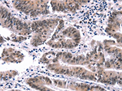
The image on the left is immunohistochemistry of paraffin-embedded Human colon cancer tissue using CSB-PA993829(MYF5 Antibody) at dilution 1/15, on the right is treated with synthetic peptide. (Original magnification: x200)
 at dilution 1/15, on the right is treated with synthetic peptide. (Original magnification: x200)")
The image on the left is immunohistochemistry of paraffin-embedded Human colon cancer tissue using CSB-PA993829(MYF5 Antibody) at dilution 1/15, on the right is treated with synthetic peptide. (Original magnification: x200)
MYF5 Antibody
CSB-PA993829
ApplicationsELISA, ImmunoHistoChemistry
Product group Antibodies
TargetMYF5
Overview
- SupplierCusabio
- Product NameMYF5 Antibody
- Delivery Days Customer20
- ApplicationsELISA, ImmunoHistoChemistry
- CertificationResearch Use Only
- ClonalityPolyclonal
- ConjugateUnconjugated
- Gene ID4617
- Target nameMYF5
- Target descriptionmyogenic factor 5
- Target synonymsbHLHc2; class C basic helix-loop-helix protein 2; EORVA; myf-5; myogenic factor 5
- HostRabbit
- IsotypeIgG
- Protein IDP13349
- Protein NameMyogenic factor 5
- Scientific DescriptionMyogenic factor 5 is a protein that in humans is encoded by the MYF5 gene. It is a protein with a key role in regulating muscle differentiation or myogenesis. Without Myf5 and MyoD, myogenic cells will fail to progress normally during the determination stage of myogenesis. Myf5 belongs to a family of proteins known as myogenic regulatory factors (MRFs). These bHLH (basic helix loop helix) transcription factors act sequentially in myogenic differentiation. MRF family members include Myf5, MyoD (Myf3), myogenin, and MRF4 (Myf6).
- Storage Instruction-20°C or -80°C
- UNSPSC12352203
Related products

Product group Antibodies
Myf5 Polyclonal AntibodyCAC11721
ApplicationsWestern Blot, ELISA
TargetMYF5
- SizePrice
Product group Antibodies
Anti-MYF5 AntibodyA96380
ApplicationsImmunoFluorescence, Western Blot, ELISA, ImmunoHistoChemistry
- SizePrice
Product group Antibodies
MYF5 Polyclonal AntibodyBS-6936R
ApplicationsImmunoFluorescence, ELISA, ImmunoCytoChemistry, ImmunoHistoChemistry, ImmunoHistoChemistry Frozen, ImmunoHistoChemistry Paraffin
TargetMYF5
- SizePrice
Product group Antibodies

MYF5 / MYF 5 AntibodyLS-C670731
ApplicationsWestern Blot, ELISA
TargetMYF5
- SizePrice
Product group Antibodies
Anti-Myf5 Antibody Picoband(r)A04040-1-CARRIER-FREE
ApplicationsWestern Blot, ELISA
TargetMYF5
- SizePrice