

MYOD / MYOD1 Antibody (clone 5.8A)
LS-C391653
ApplicationsFlow Cytometry, ImmunoFluorescence, ImmunoHistoChemistry, ImmunoHistoChemistry Paraffin
Product group Antibodies
ReactivityHuman, Mouse, Rat
TargetMYOD1
Overview
- SupplierLifeSpan BioSciences
- Product NameMYOD / MYOD1 Antibody (clone 5.8A)
- Delivery Days Customer23
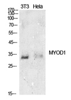
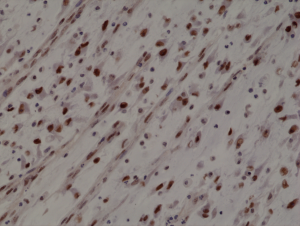
- Application Supplier NoteRecognizes a phosphor-protein of 45kDa, identified as MyoD1. The epitope of this mAb maps between amino acid 180-189 in the C-terminal of mouse MyoD1 protein. It does not cross react with myogenin, Myf5, or Myf6. Antibody to MyoD1 labels the nuclei of myoblasts in developing muscle tissues. MyoD1 is not detected in normal adult tissue, but is highly expressed in the tumor cell nuclei of rhabdomyosarcomas. Occasionally nuclear expression of MyoD1 is seen in ectomesenchymoma and a subset of Wilm s tumors. Weak cytoplasmic staining is observed in several non-muscle tissues, including glandular epithelium and also in rhabdomyosarcomas, neuroblastomas, Ewing s sarcomas and alveolar soft part sarcomas. The optimal dilution of the MyoD1 antibody for each application should be determined by the researcher. Staining of formalin-fixed tissues requires boiling tissue sections in 10mM Tris with 1mM EDTA, pH 9.0, for 10-20 min followed by cooling at RT for 20 minutes.. Flo (0.5 - 1 µg/10E6 cells), IF (0.5 - 1 µg/ml), IHC-P (0.5 - 1 µg/ml) Recognizes a phosphor-protein of 45kDa, identified as MyoD1. The epitope of this mAb maps between amino acid 180-189 in the C-terminal of mouse MyoD1 protein. It does not cross react with myogenin, Myf5, or Myf6. Antibody to MyoD1 labels the nuclei of myoblasts in developing muscle tissues. MyoD1 is not detected in normal adult tissue, but is highly expressed in the tumor cell nuclei of rhabdomyosarcomas. Occasionally nuclear expression of MyoD1 is seen in ectomesenchymoma and a subset of Wilm s tumors. Weak cytoplasmic staining is observed in several non-muscle tissues, including glandular epithelium and also in rhabdomyosarcomas, neuroblastomas, Ewing s sarcomas and alveolar soft part sarcomas. The optimal dilution of the MyoD1 antibody for each application should be determined by the researcher. Staining of formalin-fixed tissues requires boiling tissue sections in 10mM Tris with 1mM EDTA, pH 9.0, for 10-20 min followed by cooling at RT for 20 minutes.
- ApplicationsFlow Cytometry, ImmunoFluorescence, ImmunoHistoChemistry, ImmunoHistoChemistry Paraffin
- CertificationResearch Use Only
- ClonalityMonoclonal
- Clone ID5.8A
- Concentration0.2 mg/ml
- ConjugateUnconjugated
- Estimated Purity...
- Gene ID4654
- Target nameMYOD1
- Target descriptionmyogenic differentiation 1
- Target synonymsCMYO17, CMYP17, MYF3, MYOD, MYODRIF, PUM, bHLHc1, myoblast determination protein 1, class C basic helix-loop-helix protein 1, myf-3, myogenic factor 3
- HostMouse
- IsotypeIgG1
- ReactivityHuman, Mouse, Rat
- Storage Instruction2°C to 8°C
- UNSPSC12352203
Related products
Product group Antibodies
MyoD1/Myf3 Polyclonal AntibodyBS-23809R
ApplicationsWestern Blot, ImmunoHistoChemistry, ImmunoHistoChemistry Paraffin
ReactivityBovine, Canine, Equine, Human, Mouse, Porcine, Rat, Sheep
TargetMYOD1
- SizePrice
Product group Antibodies
MYOD1 Polyclonal AntibodyCAC14646
ApplicationsWestern Blot, ELISA
ReactivityMouse
TargetMYOD1
- SizePrice
Product group Antibodies
Anti-MyoD1 Antibody188-11549
ApplicationsFlow Cytometry
ReactivityChicken, Human, Mouse, Rat
TargetMYOD1
- SizePrice
Product group Antibodies
Anti-MYOD1 AntibodyA97396
ApplicationsWestern Blot, ELISA
ReactivityHuman, Mouse, Rat
- SizePrice
Product group Antibodies

Anti-MYOD1 [6F4-2D7]Ab03307-10.0
ApplicationsWestern Blot, ELISA, ImmunoHistoChemistry
ReactivityHuman
TargetMYOD1
- SizePrice
Product group Antibodies
References
MyoD1 antibody [HL1372]GTX636812
ApplicationsImmunoFluorescence, Western Blot, ImmunoCytoChemistry, ImmunoHistoChemistry, ImmunoHistoChemistry Paraffin
ReactivityHuman, Mouse, Rat
TargetMYOD1
- SizePrice
Product group Antibodies
anti-MyoD1 (human), Rabbit Monoclonal (RM369)REV-31-1255-00
ApplicationsWestern Blot, ImmunoHistoChemistry
ReactivityHuman
TargetMYOD1
- SizePrice
Product group Antibodies

MYOD / MYOD1 Antibody (clone ABT-MYOD1)LS-C744545
ApplicationsWestern Blot, ImmunoHistoChemistry
ReactivityHuman
TargetMYOD1
- SizePrice