

Various tissue extracts (50 μg) were separated by 7.5% SDS-PAGE, and the membrane was blotted with NMDAR1 antibody (GTX133097) diluted at 1:1000. The HRP-conjugated anti-rabbit IgG antibody (GTX213110-01) was used to detect the primary antibody.
![NMDAR1 antibody detects NMDAR1 protein at synaptic vesicles by immunofluorescent analysis. Sample: DIV9 rat E18 primary cortical neurons were fixed in 4% paraformaldehyde at RT for 15 min. Green: NMDAR1 protein stained by NMDAR1 antibody (GTX133097) diluted at 1:500. Red: beta Tubulin 3/ Tuj1, stained by beta Tubulin 3/ Tuj1 antibody [GT11710] (GTX631836) diluted at 1:500. Blue:Fluoroshield with DAPI (GTX30920).](https://www.genetex.com/upload/website/prouct_img/normal/GTX133097/GTX133097_42508_20170503_IFA_R_w_23060523_608.webp "NMDAR1 antibody detects NMDAR1 protein at synaptic vesicles by immunofluorescent analysis. Sample: DIV9 rat E18 primary cortical neurons were fixed in 4% paraformaldehyde at RT for 15 min. Green: NMDAR1 protein stained by NMDAR1 antibody (GTX133097) diluted at 1:500. Red: beta Tubulin 3/ Tuj1, stained by beta Tubulin 3/ Tuj1 antibody [GT11710] (GTX631836) diluted at 1:500. Blue:Fluoroshield with DAPI (GTX30920).")
 diluted at 1:400.
Antigen Retrieval: Citrate buffer, pH 6.0, 15 min")
![NMDAR1 antibody detects NMDAR1 protein expression by immunohistochemical analysis. Sample: Frozen sectioned adult mouse retina. Green: NMDAR1 protein stained by NMDAR1 antibody (GTX133097) diluted at 1:250. Red: beta Tubulin 3/ TUJ1, stained by beta Tubulin 3/ TUJ1 antibody [GT11710] (GTX631836) diluted at 1:250. Blue: Fluoroshield with DAPI (GTX30920).](https://www.genetex.com/upload/website/prouct_img/normal/GTX133097/GTX133097_42508_20170220_IHC-Fr_M_w_23060523_981.webp "NMDAR1 antibody detects NMDAR1 protein expression by immunohistochemical analysis. Sample: Frozen sectioned adult mouse retina. Green: NMDAR1 protein stained by NMDAR1 antibody (GTX133097) diluted at 1:250. Red: beta Tubulin 3/ TUJ1, stained by beta Tubulin 3/ TUJ1 antibody [GT11710] (GTX631836) diluted at 1:250. Blue: Fluoroshield with DAPI (GTX30920).")
![NMDAR1 antibody detects NMDAR1 protein by immunofluorescent analysis. Sample: DIV10 rat E18 primary cortical neuron cells were fixed in 4% paraformaldehyde at RT for 15 min. Green: NMDAR1 stained by NMDAR1 antibody (GTX133097) diluted at 1:500. Red: Tau, stained by Tau antibody [GT287] (GTX634809) diluted at 1:500. Blue: Fluoroshield with DAPI (GTX30920).](https://www.genetex.com/upload/website/prouct_img/normal/GTX133097/GTX133097_43712_20191125_ICC_IF_R_w_23060523_808.webp "NMDAR1 antibody detects NMDAR1 protein by immunofluorescent analysis. Sample: DIV10 rat E18 primary cortical neuron cells were fixed in 4% paraformaldehyde at RT for 15 min. Green: NMDAR1 stained by NMDAR1 antibody (GTX133097) diluted at 1:500. Red: Tau, stained by Tau antibody [GT287] (GTX634809) diluted at 1:500. Blue: Fluoroshield with DAPI (GTX30920).")
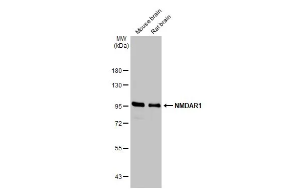
Various tissue extracts (50 μg) were separated by 7.5% SDS-PAGE, and the membrane was blotted with NMDAR1 antibody (GTX133097) diluted at 1:1000. The HRP-conjugated anti-rabbit IgG antibody (GTX213110-01) was used to detect the primary antibody.
NMDAR1 antibody
GTX133097
ApplicationsImmunoFluorescence, Western Blot, ImmunoCytoChemistry, ImmunoHistoChemistry, ImmunoHistoChemistry Frozen, ImmunoHistoChemistry Paraffin
Product group Antibodies
ReactivityCanine, Mouse, Rat
TargetGrin1
Overview
- SupplierGeneTex
- Product NameNMDAR1 antibody
- Delivery Days Customer9
- Application Supplier NoteWB: 1:500-1:3000. ICC/IF: 1:100-1:1000. IHC-P: 1:100-1:1000. IHC-Fr: 1:100-1:1000. *Optimal dilutions/concentrations should be determined by the researcher.Not tested in other applications.
- ApplicationsImmunoFluorescence, Western Blot, ImmunoCytoChemistry, ImmunoHistoChemistry, ImmunoHistoChemistry Frozen, ImmunoHistoChemistry Paraffin
- CertificationResearch Use Only
- ClonalityPolyclonal
- Concentration1.37 mg/ml
- ConjugateUnconjugated
- Gene ID14810
- Target nameGrin1
- Target descriptionglutamate receptor, ionotropic, NMDA1 (zeta 1)
- Target synonymsGluN1, GluRdelta1, GluRzeta1, M100174, NMD-R1, NMDAR1, NR1, Nmdar, Rgsc174, glutamate receptor ionotropic, NMDA 1, N-methyl-D-aspartate receptor subunit NR1, glutamate [NMDA] receptor subunit zeta-1
- HostRabbit
- IsotypeIgG
- Protein IDP35438
- Protein NameGlutamate receptor ionotropic, NMDA 1
- Scientific DescriptionNMDA receptor subtype of glutamate-gated ion channels possesses high calcium permeability and voltage-dependent sensitivity to magnesium. Modulated by glycine. This protein plays a key role in synaptic plasticity, synaptogenesis, excitotoxicity, memory acquisition and learning. It mediates neuronal functions in glutamate neurotransmission. Is involved in the cell surface targeting of NMDA receptors.
- ReactivityCanine, Mouse, Rat
- Storage Instruction-20°C or -80°C,2°C to 8°C
- UNSPSC41116161