

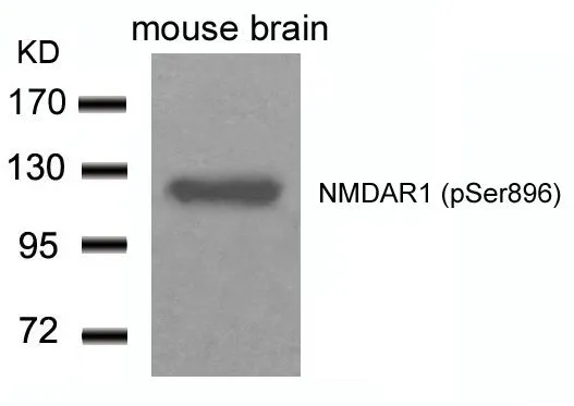
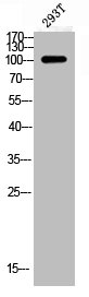
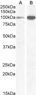
WB analysis of extracts from mouse brain tissue using GTX50168 NMDAR1 (phospho Ser896) antibody.
WB analysis of extracts from mouse brain tissue using GTX50168 NMDAR1 (phospho Ser896) antibody.
NMDAR1 (phospho Ser896) antibody
GTX50168
ApplicationsWestern Blot
Product group Antibodies
ReactivityHuman, Mouse
TargetGRIN1
Overview
- SupplierGeneTex
- Product NameNMDAR1 (phospho Ser896) antibody
- Delivery Days Customer9
- Application Supplier NoteWB: 1:500-1:1000. *Optimal dilutions/concentrations should be determined by the researcher.Not tested in other applications.
- ApplicationsWestern Blot
- CertificationResearch Use Only
- ClonalityPolyclonal
- Concentration1 mg/ml
- ConjugateUnconjugated
- Gene ID2902
- Target nameGRIN1
- Target descriptionglutamate ionotropic receptor NMDA type subunit 1
- Target synonymsDEE101, GluN1, MRD8, NDHMSD, NDHMSR, NMD-R1, NMDA1, NMDAR1, NR1, hNR1, glutamate receptor ionotropic, NMDA 1, N-methyl-D-aspartate receptor channel, subunit zeta-1, N-methyl-D-aspartate receptor subunit NR1, glutamate [NMDA] receptor subunit zeta-1, glutamate receptor, ionotropic, N-methyl D-aspartate 1, putative NMDtranscript(altAcc_e2)
- HostRabbit
- IsotypeIgG
- Protein IDQ05586
- Protein NameGlutamate receptor ionotropic, NMDA 1
- Scientific DescriptionThe protein encoded by this gene is a critical subunit of N-methyl-D-aspartate receptors, members of the glutamate receptor channel superfamily which are heteromeric protein complexes with multiple subunits arranged to form a ligand-gated ion channel. These subunits play a key role in the plasticity of synapses, which is believed to underlie memory and learning. Cell-specific factors are thought to control expression of different isoforms, possibly contributing to the functional diversity of the subunits. Alternatively spliced transcript variants have been described. [provided by RefSeq, Jul 2008]
- ReactivityHuman, Mouse
- Storage Instruction-20°C or -80°C,2°C to 8°C
- UNSPSC41116161
Datasheet
Related products
Product group Antibodies
Anti-NMDAR1 AntibodyA83429
ApplicationsImmunoFluorescence, ELISA
ReactivityHuman, Mouse
- SizePrice
Product group Antibodies
Anti-GRIN1 Antibody144-66462
ApplicationsImmunoFluorescence, Western Blot, ImmunoHistoChemistry
ReactivityHuman, Mouse, Rat
TargetGRIN1
- SizePrice
Product group Antibodies
Anti-NMDAR1/GRIN1 Antibody Picoband(r)A01808-CARRIER-FREE
ApplicationsWestern Blot
ReactivityHuman, Mouse, Rat
TargetGRIN1
- SizePrice
Product group Antibodies
References
NMDAR1 Polyclonal AntibodyBS-2175R
ApplicationsImmunoFluorescence, Western Blot, ELISA, ImmunoCytoChemistry, ImmunoHistoChemistry, ImmunoHistoChemistry Frozen, ImmunoHistoChemistry Paraffin
ReactivityCanine, Chicken, Human, Mouse, Porcine, Rabbit, Rat
TargetGRIN1
- SizePrice
Product group Antibodies
Phospho-GRIN1 (S896) AntibodyCSB-PA008967
ApplicationsWestern Blot, ELISA, ImmunoHistoChemistry
ReactivityHuman, Mouse, Rat
TargetGRIN1
- SizePrice
Product group Antibodies

ApplicationsImmunoFluorescence, ELISA
ReactivityCanine, Human, Mouse, Rat
TargetGRIN1
- SizePrice
Product group Antibodies
Grin1 Recombinant AntibodyCAC12218
ApplicationsFlow Cytometry, ELISA
TargetGRIN1
- SizePrice
Product group Antibodies

GRIN1 / NMDAR1 AntibodyLS-C402488
ApplicationsELISA, ImmunoHistoChemistry
ReactivityHuman, Mouse, Rat
TargetGRIN1
- SizePrice
Product group Antibodies
NMDAR1 antibody, InternalGTX89302
ApplicationsWestern Blot
ReactivityHuman, Rat
TargetGRIN1
- SizePrice

Product group Antibodies
Anti-GRIN1 AntibodyHPA067773
ApplicationsImmunoHistoChemistry
ReactivityHuman
TargetGRIN1
- SizePrice