

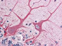
anti-NOTCH 1 antibody was diluted 1:500 to detect NOTCH 1 in human brain cerebellum tissue. Tissue was formalin fixed and paraffin embedded. No pre-treatment of sample was required. The image shows the localization of antibody as the precipitated red signal, with a hematoxylin purple nuclear counter stain.
anti-NOTCH 1 antibody was diluted 1:500 to detect NOTCH 1 in human brain cerebellum tissue. Tissue was formalin fixed and paraffin embedded. No pre-treatment of sample was required. The image shows the localization of antibody as the precipitated red signal, with a hematoxylin purple nuclear counter stain.
NOTCH1 Rabbit Polyclonal Antibody

TA319199
Overview
- SupplierOriGene
- Product NameNOTCH1 Rabbit Polyclonal Antibody
- Delivery Days Customer14
- ApplicationsImmunoHistoChemistry
- CertificationResearch Use Only
- ClonalityPolyclonal
- Gene ID4851
- Target nameNOTCH1
- Target descriptionnotch receptor 1
- Target synonymsAOS5; AOVD1; hN1; neurogenic locus notch homolog protein 1; notch 1; Notch homolog 1, translocation-associated; TAN1; translocation-associated notch protein TAN-1
- HostRabbit
- IsotypeIgG
- Protein IDP46531
- Protein NameNeurogenic locus notch homolog protein 1
- Scientific DescriptionRabbit polyclonal anti-Notch 1 antibody
- ReactivityHuman
- Storage Instruction-20°C
- UNSPSC12352203