

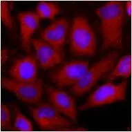
Immunofluorescence of human HeLa cells stained with monoclonal anti-human OAT anitbody (1:500) with Texas Red (Red). Nucleus was stained by Hoechst 33342 (Blue).
 were resolved by SDS-PAGE, transferred to NC membrane and probed with anti-human OAT (1:1000). Proteins were visualized using a goat anti-mouse secondary antibody conjugated to HRP and an ECL detection system.")
Immunofluorescence of human HeLa cells stained with monoclonal anti-human OAT anitbody (1:500) with Texas Red (Red). Nucleus was stained by Hoechst 33342 (Blue).
OAT antibody [AT23A2]
GTX50004
ApplicationsFlow Cytometry, ImmunoFluorescence, Western Blot, ELISA, ImmunoCytoChemistry
Product group Antibodies
ReactivityHuman, Mouse
TargetOAT
Overview
- SupplierGeneTex
- Product NameOAT antibody [AT23A2]
- Delivery Days Customer9
- ApplicationsFlow Cytometry, ImmunoFluorescence, Western Blot, ELISA, ImmunoCytoChemistry
- CertificationResearch Use Only
- ClonalityMonoclonal
- Clone IDAT23A2
- Concentration1 mg/ml
- ConjugateUnconjugated
- Gene ID4942
- Target nameOAT
- Target descriptionornithine aminotransferase
- Target synonymsGACR, HOGA, OATASE, OKT, ornithine aminotransferase, mitochondrial, gyrate atrophy, ornithine delta-aminotransferase, ornithine-oxo-acid aminotransferase, testicular tissue protein Li 130
- HostMouse
- IsotypeIgG1
- Protein IDP04181
- Protein NameOrnithine aminotransferase, mitochondrial
- Scientific DescriptionThis gene encodes the mitochondrial enzyme ornithine aminotransferase, which is a key enzyme in the pathway that converts arginine and ornithine into the major excitatory and inhibitory neurotransmitters glutamate and GABA. Mutations that result in a deficiency of this enzyme cause the autosomal recessive eye disease Gyrate Atrophy. Alternatively spliced transcript variants encoding different isoforms have been described. Related pseudogenes have been defined on the X chromosome. [provided by RefSeq, Jan 2010]
- ReactivityHuman, Mouse
- Storage Instruction-20°C or -80°C,2°C to 8°C
- UNSPSC41116161
Datasheet
Related products
Product group Antibodies
OAT AntibodyCSB-PA020180
ApplicationsWestern Blot, ELISA
ReactivityHuman, Mouse, Rat
TargetOAT
- SizePrice
Product group Antibodies
Anti-OAT Antibody144-06235
ApplicationsImmunoPrecipitation, Western Blot
ReactivityHuman, Mouse, Rat
TargetOAT
- SizePrice

Product group Antibodies
Anti-ornithine aminotransferase/OAT Antibody Picoband(r)A01126-2-CARRIER-FREE
ApplicationsFlow Cytometry, Western Blot, ELISA, ImmunoHistoChemistry
ReactivityHuman, Mouse, Rat
TargetOAT
- SizePrice
Product group Antibodies
ApplicationsImmunoFluorescence, Western Blot, ELISA
ReactivityHuman
- SizePrice

Product group Antibodies
Anti-OAT AntibodyHPA040098
ApplicationsWestern Blot, ImmunoHistoChemistry
ReactivityHuman
TargetOAT
- SizePrice
Product group Antibodies

OAT AntibodyLS-C334577
ApplicationsWestern Blot, ImmunoHistoChemistry
ReactivityHuman, Mouse, Rat
TargetOAT
- SizePrice
![OAT antibody [HL2087] detects OAT protein at cytoplasm by immunohistochemical analysis. Sample: Paraffin-embedded mouse adrenal gland. OAT stained by OAT antibody [HL2087] (GTX637995) diluted at 1:100. Antigen Retrieval: Citrate buffer, pH 6.0, 15 min](https://www.genetex.com/upload/website/prouct_img/normal/GTX637995/GTX637995_T-44900_20230113_IHC-P_M_23013122_615.webp)
Product group Antibodies
OAT antibody [HL2087]GTX637995
ApplicationsImmunoFluorescence, Western Blot, ImmunoCytoChemistry, ImmunoHistoChemistry, ImmunoHistoChemistry Paraffin
ReactivityHuman, Mouse, Rat
TargetOAT
- SizePrice
![OAT antibody [HL2088] detects OAT protein at cytoplasm by immunohistochemical analysis. Sample: Paraffin-embedded rat duodenum. OAT stained by OAT antibody [HL2088] (GTX637996) diluted at 1:100. Antigen Retrieval: Citrate buffer, pH 6.0, 15 min](https://www.genetex.com/upload/website/prouct_img/normal/GTX637996/GTX637996_T-44900_20230113_IHC-P_R_23013122_962.webp)
Product group Antibodies
OAT antibody [HL2088]GTX637996
ApplicationsImmunoFluorescence, Western Blot, ImmunoCytoChemistry, ImmunoHistoChemistry, ImmunoHistoChemistry Paraffin
ReactivityHuman, Mouse, Rat
TargetOAT
- SizePrice
![Non-transfected (–) and transfected (+) 293T whole cell extracts (30 μg) were separated by 10% SDS-PAGE, and the membrane was blotted with OAT antibody [N1C3] (GTX103893) diluted at 1:5000. The HRP-conjugated anti-rabbit IgG antibody (GTX213110-01) was used to detect the primary antibody.](https://www.genetex.com/upload/website/prouct_img/normal/GTX103893/GTX103893_43131_20180817_WB_B_w_23060120_171.webp)
Product group Antibodies
OAT antibody [N1C3]GTX103893
ApplicationsImmunoFluorescence, Western Blot, ImmunoCytoChemistry, ImmunoHistoChemistry, ImmunoHistoChemistry Paraffin
ReactivityHuman, Mouse
TargetOAT
- SizePrice