

![OLIG2 (Marker of Glial Brain Tumors) (OLIG2/2400), CF405S conjugate, 0.1mg/mL [26628-22-8]](https://biotium.com/wp-content/uploads/2018/10/BNUB2400-1.jpeg "OLIG2 (Marker of Glial Brain Tumors) (OLIG2/2400), CF405S conjugate, 0.1mg/mL [26628-22-8]")
![OLIG2 (Marker of Glial Brain Tumors) (OLIG2/2400), CF405S conjugate, 0.1mg/mL [26628-22-8]](https://biotium.com/wp-content/uploads/2018/10/BNUB2400-2.jpeg "OLIG2 (Marker of Glial Brain Tumors) (OLIG2/2400), CF405S conjugate, 0.1mg/mL [26628-22-8]")
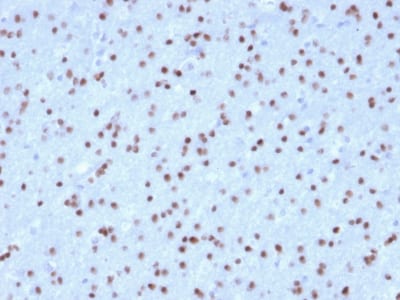
OLIG2 (Marker of Glial Brain Tumors) (OLIG2/2400), CF405S conjugate, 0.1mg/mL [26628-22-8]
BNC042400
ApplicationsImmunoHistoChemistry, ImmunoHistoChemistry Paraffin
Product group Antibodies
ReactivityBovine, Human, Mouse
TargetOLIG2
Overview
- SupplierBiotium
- Product NameOLIG2 (Marker of Glial Brain Tumors) (OLIG2/2400), CF405S conjugate, 0.1mg/mL
- Delivery Days Customer9
- ApplicationsImmunoHistoChemistry, ImmunoHistoChemistry Paraffin
- CertificationResearch Use Only
- ClonalityMonoclonal
- Clone IDOLIG2/2400
- Concentration0.1 mg/ml
- ConjugateOther Conjugate
- Gene ID10215
- Target nameOLIG2
- Target descriptionoligodendrocyte transcription factor 2
- Target synonymsbasic domain, helix-loop-helix protein, class B, 1; BHLHB1; bHLHe19; class B basic helix-loop-helix protein 1; class E basic helix-loop-helix protein 19; human protein kinase C-binding protein RACK17; OLIGO2; oligodendrocyte lineage transcription factor 2; oligodendrocyte transcription factor 2; oligodendrocyte-specific bHLH transcription factor 2; PRKCBP2; protein kinase C-binding protein 2; RACK17
- HostMouse
- IsotypeIgG1
- Protein IDQ13516
- Protein NameOligodendrocyte transcription factor 2
- Scientific DescriptionOlig2, a basic helix-loop-helix transcription factor, is involved in oligodendroglial specification. Olig2 expression has been reported in most glial tumors, such as oligodendrogliomas and astrocytomas. Although more than half of glioblastomas are positive for Olig2, expression is very weak in terms of both percentage of labeled cells and intensity. No Olig2 expression has been found in the non-glial tumors including neuro-epithelial tumors, ependymomas, sub-ependymomas, medulloblastomas, and non-neuroepithelial tumors, such as CNS lymphomas, meningiomas, schwannomas, atypical teratoid / rhabdoid tumor, and haemangioblastomas. Compared to the strong staining seen in glioma samples, a weak expression is observed in non-tumoral brain tissue (gliosis).Primary antibodies are available purified, or with a selection of fluorescent CF® Dyes and other labels. CF® Dyes offer exceptional brightness and photostability. Note: Conjugates of blue fluorescent dyes like CF®405S and CF®405M are not recommended for detecting low abundance targets, because blue dyes have lower fluorescence and can give higher non-specific background than other dye colors.
- SourceAnimal
- ReactivityBovine, Human, Mouse
- Storage Instruction2°C to 8°C
- UNSPSC12352203