

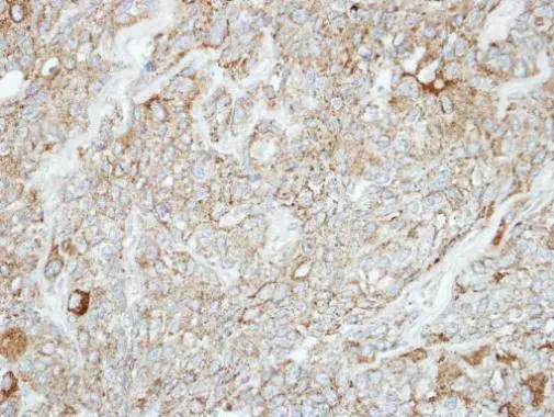
Immunohistochemical analysis of paraffin-embedded BT474 xenograft, using PCDH1(GTX114620) antibody at 1:100 dilution.
Antigen Retrieval: Citrate buffer, pH 6.0, 15 min
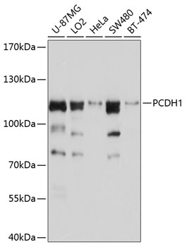
![Various whole cell extracts (30 μg) were separated by 7.5% SDS-PAGE, and the membrane was blotted with PCDH1 antibody [C2C3], C-term (GTX114620) diluted at 1:1000. The HRP-conjugated anti-rabbit IgG antibody (GTX213110-01) was used to detect the primary antibody. Corresponding RNA expression data for the same cell lines are based on Human Protein Atlas program.](https://www.genetex.com/upload/website/prouct_img/normal/GTX114620/GTX114620_40205_20231103_WB_TPM_watermark_23110819_913.webp "Various whole cell extracts (30 μg) were separated by 7.5% SDS-PAGE, and the membrane was blotted with PCDH1 antibody [C2C3], C-term (GTX114620) diluted at 1:1000. The HRP-conjugated anti-rabbit IgG antibody (GTX213110-01) was used to detect the primary antibody. Corresponding RNA expression data for the same cell lines are based on Human Protein Atlas program.")
Immunohistochemical analysis of paraffin-embedded BT474 xenograft, using PCDH1(GTX114620) antibody at 1:100 dilution.
Antigen Retrieval: Citrate buffer, pH 6.0, 15 min
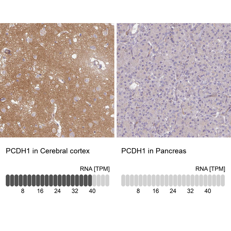
PCDH1 antibody [C2C3], C-term
GTX114620
ApplicationsWestern Blot, ImmunoHistoChemistry, ImmunoHistoChemistry Paraffin
Product group Antibodies
ReactivityHuman, Mouse
TargetPCDH1
Overview
- SupplierGeneTex
- Product NamePCDH1 antibody [C2C3], C-term
- Delivery Days Customer9
- Application Supplier NoteWB: 1:500-1:3000. IHC-P: 1:100-1:1000. *Optimal dilutions/concentrations should be determined by the researcher.Not tested in other applications.
- ApplicationsWestern Blot, ImmunoHistoChemistry, ImmunoHistoChemistry Paraffin
- CertificationResearch Use Only
- ClonalityPolyclonal
- Concentration0.69 mg/ml
- ConjugateUnconjugated
- Gene ID5097
- Target namePCDH1
- Target descriptionprotocadherin 1
- Target synonymsPC42, PCDH42, protocadherin-1, cadherin-like 1, cadherin-like protein 1, protocadherin 42
- HostRabbit
- IsotypeIgG
- Protein IDQ08174
- Protein NameProtocadherin-1
- Scientific DescriptionThis gene belongs to the protocadherin subfamily within the cadherin superfamily. The encoded protein is a membrane protein found at cell-cell boundaries. It is involved in neural cell adhesion, suggesting a possible role in neuronal development. The protein includes an extracelllular region, containing 7 cadherin-like domains, a transmembrane region and a C-terminal cytoplasmic region. Cells expressing the protein showed cell aggregation activity. Alternative splicing occurs in this gene. [provided by RefSeq]
- ReactivityHuman, Mouse
- Storage Instruction-20°C or -80°C,2°C to 8°C
- UNSPSC41116161
Datasheet
Related products

Product group Antibodies
ApplicationsWestern Blot
ReactivityHuman, Mouse
TargetPCDH1
- SizePrice
Product group Antibodies
Anti-PCDH1 AntibodyA11270
ApplicationsWestern Blot
ReactivityHuman, Mouse
- SizePrice
Product group Antibodies
Anti-PCDH1 Antibody144-10234
ApplicationsWestern Blot
ReactivityHuman, Mouse
TargetPCDH1
- SizePrice
Product group Antibodies
ApplicationsImmunoPrecipitation, Western Blot, ImmunoCytoChemistry, ImmunoHistoChemistry
ReactivityMouse, Rat
TargetPCDH1
- SizePrice
Product group Antibodies
PCDH1 AntibodyCSB-PA2106ESR2HU
ApplicationsWestern Blot, ELISA, ImmunoHistoChemistry
ReactivityHuman, Mouse
TargetPCDH1
- SizePrice
Product group Antibodies
PCDH1 antibody [N1N3]GTX103806
ApplicationsImmunoFluorescence, Western Blot, ImmunoCytoChemistry
ReactivityHuman
TargetPCDH1
- SizePrice
Product group Antibodies

PCDH1 / PCD1 AntibodyLS-C496942
ApplicationsWestern Blot
ReactivityHuman, Mouse
TargetPCDH1
- SizePrice
Product group Antibodies
Anti-PCDH1 AntibodyHPA050538
ApplicationsImmunoHistoChemistry
ReactivityHuman
TargetPCDH1
- SizePrice