

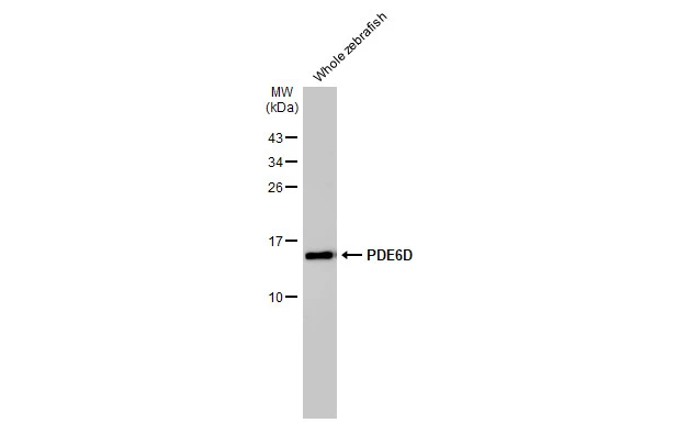
Whole zebrafish extract (30 μg) was separated by 15% SDS-PAGE, and the membrane was blotted with PDE6D antibody [HL1359] (GTX636799) diluted at 1:1000. The HRP-conjugated anti-rabbit IgG antibody (GTX213110-01) was used to detect the primary antibody.
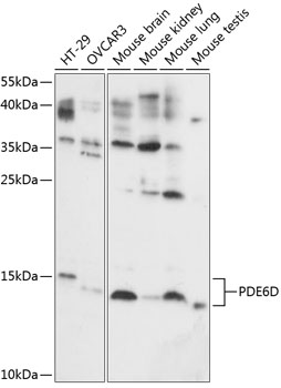
![Various whole cell extracts (30 μg) were separated by 15% SDS-PAGE, and the membrane was blotted with PDE6D antibody [HL1359] (GTX636799) diluted at 1:1000. The HRP-conjugated anti-rabbit IgG antibody (GTX213110-01) was used to detect the primary antibody.](https://www.genetex.com/upload/website/prouct_img/normal/GTX636799/GTX636799_44648_20221223_WB_D_C_22122722_604.webp "Various whole cell extracts (30 μg) were separated by 15% SDS-PAGE, and the membrane was blotted with PDE6D antibody [HL1359] (GTX636799) diluted at 1:1000. The HRP-conjugated anti-rabbit IgG antibody (GTX213110-01) was used to detect the primary antibody.")
![PDE6D antibody [HL1359] detects PDE6D protein at cytoplasm by immunohistochemical analysis. Sample: Paraffin-embedded cat colon. PDE6D stained by PDE6D antibody [HL1359] (GTX636799) diluted at 1:100. Antigen Retrieval: Citrate buffer, pH 6.0, 15 min](https://www.genetex.com/upload/website/prouct_img/normal/GTX636799/GTX636799_44648_20230331_IHC-P_Cat_23032819_237.webp "PDE6D antibody [HL1359] detects PDE6D protein at cytoplasm by immunohistochemical analysis. Sample: Paraffin-embedded cat colon. PDE6D stained by PDE6D antibody [HL1359] (GTX636799) diluted at 1:100. Antigen Retrieval: Citrate buffer, pH 6.0, 15 min")
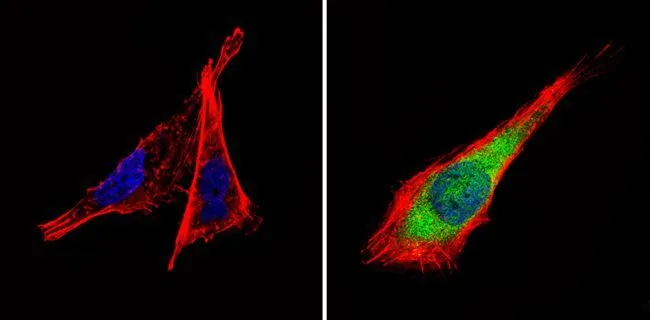
![PDE6D antibody [HL1359] detects PDE6D protein at nucleus by immunofluorescent analysis. Sample: MOLT-4 cells were fixed in 4% paraformaldehyde at RT for 15 min. Green: PDE6D stained by PDE6D antibody [HL1359] (GTX636799) diluted at 1:500. Blue: Fluoroshield with DAPI (GTX30920).](https://www.genetex.com/upload/website/prouct_img/normal/GTX636799/GTX636799_44648_20230512_ICC_IF_23060622_954.webp "PDE6D antibody [HL1359] detects PDE6D protein at nucleus by immunofluorescent analysis. Sample: MOLT-4 cells were fixed in 4% paraformaldehyde at RT for 15 min. Green: PDE6D stained by PDE6D antibody [HL1359] (GTX636799) diluted at 1:500. Blue: Fluoroshield with DAPI (GTX30920).")
![Mouse tissue extract (50 μg) was separated by 15% SDS-PAGE, and the membrane was blotted with PDE6D antibody [HL1359] (GTX636799) diluted at 1:1000. The HRP-conjugated anti-rabbit IgG antibody (GTX213110-01) was used to detect the primary antibody.](https://www.genetex.com/upload/website/prouct_img/normal/GTX636799/GTX636799_T-44599_20220311_WB_M_eye_w_23061202_140.webp "Mouse tissue extract (50 μg) was separated by 15% SDS-PAGE, and the membrane was blotted with PDE6D antibody [HL1359] (GTX636799) diluted at 1:1000. The HRP-conjugated anti-rabbit IgG antibody (GTX213110-01) was used to detect the primary antibody.")
![PDE6D antibody [HL1359] detects PDE6D protein at cytoplasm by immunohistochemical analysis. Sample: Paraffin-embedded mouse eye. Green: PDE6D stained by PDE6D antibody [HL1359] (GTX636799) diluted at 1:100. Red: beta Tubulin 3/ Tuj1, a neural marker, stained by beta Tubulin 3/ Tuj1 antibody [GT11710] (GTX631836) diluted at 1:500. Blue: Fluoroshield with DAPI (GTX30920). Antigen Retrieval: Citrate buffer, pH 6.0, 15 min](https://www.genetex.com/upload/website/prouct_img/normal/GTX636799/GTX636799_T-44599_20220415_IHC-P_M_w_23061202_615.webp "PDE6D antibody [HL1359] detects PDE6D protein at cytoplasm by immunohistochemical analysis. Sample: Paraffin-embedded mouse eye. Green: PDE6D stained by PDE6D antibody [HL1359] (GTX636799) diluted at 1:100. Red: beta Tubulin 3/ Tuj1, a neural marker, stained by beta Tubulin 3/ Tuj1 antibody [GT11710] (GTX631836) diluted at 1:500. Blue: Fluoroshield with DAPI (GTX30920). Antigen Retrieval: Citrate buffer, pH 6.0, 15 min")
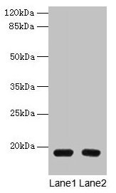
![Whole cell extract (30 μg) was separated by 15% SDS-PAGE, and the membrane was blotted with PDE6D antibody [HL1359] (GTX636799) diluted at 1:1000. The HRP-conjugated anti-rabbit IgG antibody (GTX213110-01) was used to detect the primary antibody.](https://www.genetex.com/upload/website/prouct_img/normal/GTX636799/GTX636799_44648_20220506_WB_w_23061202_156.webp "Whole cell extract (30 μg) was separated by 15% SDS-PAGE, and the membrane was blotted with PDE6D antibody [HL1359] (GTX636799) diluted at 1:1000. The HRP-conjugated anti-rabbit IgG antibody (GTX213110-01) was used to detect the primary antibody.")
![PDE6D antibody [HL1359] detects PDE6D protein at cytoplasm by immunohistochemical analysis. Sample: Paraffin-embedded mouse intestine. PDE6D stained by PDE6D antibody [HL1359] (GTX636799) diluted at 1:100. Antigen Retrieval: Citrate buffer, pH 6.0, 15 min](https://www.genetex.com/upload/website/prouct_img/normal/GTX636799/GTX636799_44648_20220520_IHC-P_M_w_23061202_895.webp "PDE6D antibody [HL1359] detects PDE6D protein at cytoplasm by immunohistochemical analysis. Sample: Paraffin-embedded mouse intestine. PDE6D stained by PDE6D antibody [HL1359] (GTX636799) diluted at 1:100. Antigen Retrieval: Citrate buffer, pH 6.0, 15 min")
![PDE6D antibody [HL1359] detects PDE6D protein at cytoplasm by immunohistochemical analysis. Sample: Paraffin-embedded rat colon. PDE6D stained by PDE6D antibody [HL1359] (GTX636799) diluted at 1:100. Antigen Retrieval: Citrate buffer, pH 6.0, 15 min](https://www.genetex.com/upload/website/prouct_img/normal/GTX636799/GTX636799_44648_20220520_IHC-P_R_w_23061202_224.webp "PDE6D antibody [HL1359] detects PDE6D protein at cytoplasm by immunohistochemical analysis. Sample: Paraffin-embedded rat colon. PDE6D stained by PDE6D antibody [HL1359] (GTX636799) diluted at 1:100. Antigen Retrieval: Citrate buffer, pH 6.0, 15 min")
![Whole Japanese medaka extract (30 μg) was separated by 15% SDS-PAGE, and the membrane was blotted with PDE6D antibody [HL1359] (GTX636799) diluted at 1:1000. The HRP-conjugated anti-rabbit IgG antibody (GTX213110-01) was used to detect the primary antibody, and the signal was developed with Trident ECL plus-Enhanced.](https://www.genetex.com/upload/website/prouct_img/normal/GTX636799/GTX636799_44648_20250815_WB_medaka_25082121_517.webp "Whole Japanese medaka extract (30 μg) was separated by 15% SDS-PAGE, and the membrane was blotted with PDE6D antibody [HL1359] (GTX636799) diluted at 1:1000. The HRP-conjugated anti-rabbit IgG antibody (GTX213110-01) was used to detect the primary antibody, and the signal was developed with Trident ECL plus-Enhanced.")
Whole zebrafish extract (30 μg) was separated by 15% SDS-PAGE, and the membrane was blotted with PDE6D antibody [HL1359] (GTX636799) diluted at 1:1000. The HRP-conjugated anti-rabbit IgG antibody (GTX213110-01) was used to detect the primary antibody.
PDE6D antibody [HL1359]
GTX636799
ApplicationsImmunoFluorescence, Western Blot, ImmunoCytoChemistry, ImmunoHistoChemistry, ImmunoHistoChemistry Paraffin
Product group Antibodies
ReactivityCanine, Feline, Human, Mouse, Rat, Zebra Fish
TargetPDE6D
Overview
- SupplierGeneTex
- Product NamePDE6D antibody [HL1359]
- Delivery Days Customer9
- Application Supplier NoteWB: 1:500-1:3000. *Optimal dilutions/concentrations should be determined by the researcher.Not tested in other applications.
- ApplicationsImmunoFluorescence, Western Blot, ImmunoCytoChemistry, ImmunoHistoChemistry, ImmunoHistoChemistry Paraffin
- CertificationResearch Use Only
- ClonalityMonoclonal
- Clone IDHL1359
- Concentration1 mg/ml
- ConjugateUnconjugated
- Gene ID5147
- Target namePDE6D
- Target descriptionphosphodiesterase 6D
- Target synonymsJBTS22, PDED, retinal rod rhodopsin-sensitive cGMP 3',5'-cyclic phosphodiesterase subunit delta, GMP-PDE delta, phosphodiesterase 6D, cGMP-specific, rod, delta, protein p17
- HostRabbit
- IsotypeIgG
- Protein IDO43924
- Protein NameRetinal rod rhodopsin-sensitive cGMP 3',5'-cyclic phosphodiesterase subunit delta
- Scientific DescriptionThis gene encodes the delta subunit of rod-specific photoreceptor phosphodiesterase (PDE), a key enzyme in the phototransduction cascade. A similar protein in cow functions in solubilizing membrane-bound PDE. In addition to its role in the PDE complex, the encoded protein is thought to bind to prenyl groups of proteins to target them to subcellular organelles called cilia. Mutations in this gene are associated with Joubert syndrome-22. Alternative splicing results in multiple splice variants. [provided by RefSeq, Mar 2014]
- ReactivityCanine, Feline, Human, Mouse, Rat, Zebra Fish
- Storage Instruction-20°C or -80°C,2°C to 8°C
- UNSPSC41116161
Datasheet
Related products
Product group Antibodies
PDE6D AntibodyCSB-PA526126LA01HU
ApplicationsWestern Blot, ELISA, ImmunoHistoChemistry
ReactivityHuman
TargetPDE6D
- SizePrice

Product group Antibodies
Anti-PDE6D AntibodyA06466
ApplicationsImmunoFluorescence, Western Blot, ImmunoCytoChemistry, ImmunoHistoChemistry
ReactivityHuman, Mouse, Rat
TargetPDE6D
- SizePrice
Product group Antibodies
Anti-PDE6D AntibodyA87615
ApplicationsImmunoFluorescence, Western Blot, ImmunoCytoChemistry, ImmunoHistoChemistry
ReactivityHuman, Mouse, Rat
- SizePrice
Product group Antibodies

PDE6D / PDE6 Delta AntibodyLS-C830884
ApplicationsELISA, ImmunoHistoChemistry
ReactivityHuman, Mouse
TargetPDE6D
- SizePrice

Product group Antibodies
Anti-PDE6D AntibodyHPA037433
ApplicationsWestern Blot, ImmunoHistoChemistry
ReactivityHuman
TargetPDE6D
- SizePrice
Product group Antibodies
PDE6D antibodyGTX25665
ApplicationsImmunoFluorescence, Western Blot, ImmunoCytoChemistry, ImmunoHistoChemistry, ImmunoHistoChemistry Frozen
ReactivityBovine, Human, Mouse, Sheep
TargetPDE6D
- SizePrice
Product group Antibodies
Pde6D Polyclonal AntibodyCAC07462
ApplicationsWestern Blot, ELISA, ImmunoHistoChemistry
TargetPDE6D
- SizePrice
![Whole cell extract (30 μg) was separated by 15% SDS-PAGE, and the membrane was blotted with PDE6D antibody [N1C3] (GTX109240) diluted at 1:1000. The HRP-conjugated anti-rabbit IgG antibody (GTX213110-01) was used to detect the primary antibody.](https://www.genetex.com/upload/website/prouct_img/normal/GTX109240/GTX109240_44349_20210716_WB_23052301_728.webp)
Product group Antibodies
PDE6D antibody [N1C3]GTX109240
ApplicationsImmunoFluorescence, Western Blot, ImmunoCytoChemistry, ImmunoHistoChemistry, ImmunoHistoChemistry Paraffin
ReactivityHuman, Mouse
TargetPDE6D
- SizePrice
Product group Antibodies
Anti-PDE6D Antibody144-60782
ApplicationsWestern Blot
ReactivityHuman, Mouse
TargetPDE6D
- SizePrice