

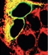
Immunohistochemistry analysis of human HEK293 cells transfected with wild type TorsinA co-stained with PDI mAb (1D3) (red). Co-localization of TorsinA and PDI proteins merge as yellow.
 Lane 3: ADI-SPP-891 Lane 4: Vero (HS) Lane 5: 3T3 (HS) Lane 6: PC-12 (HS) Lane 7: CHO-K1 (HS)")
Immunohistochemistry analysis of human HEK293 cells transfected with wild type TorsinA co-stained with PDI mAb (1D3) (red). Co-localization of TorsinA and PDI proteins merge as yellow.
PDI antibody [1D3]
GTX30716
ApplicationsImmunoFluorescence, ImmunoPrecipitation, Western Blot, ImmunoCytoChemistry, ImmunoHistoChemistry
Product group Antibodies
ReactivityBovine, Canine, Guinea Pig, Hamster, Human, Monkey, Mouse, Porcine, Rat, Sheep, Xenopus
TargetP4hb
Overview
- SupplierGeneTex
- Product NamePDI antibody [1D3]
- Delivery Days Customer9
- Application Supplier NoteFor ICC: Use at a concentration of 2-5 microg/ml. For IP: Use at a concentration of 8-12 microg/ml. For WB: Use at a concentration of 2-4 microg/ml. Predicted molecular weight: 58 kDa. Optimal dilutions/concentrations should be determined by the researcher.
- ApplicationsImmunoFluorescence, ImmunoPrecipitation, Western Blot, ImmunoCytoChemistry, ImmunoHistoChemistry
- CertificationResearch Use Only
- ClonalityMonoclonal
- Clone ID1D3
- Concentration1 mg/ml
- ConjugateUnconjugated
- Gene ID25506
- Target nameP4hb
- Target descriptionprolyl 4-hydroxylase subunit beta
- Target synonymsPDI, PDIR, protein disulfide-isomerase, Protein disulfide isomerase (Prolyl 4-hydroxylase, beta polypeptide), cellular thyroid hormone-binding protein, prolyl 4-hydroxylase, beta polypeptide
- HostMouse
- IsotypeIgG1
- Scientific DescriptionThe three dimensional structure of many extracellular proteins is stabilized by the formation of disulphide bonds. Studies suggest that a microsomal enzyme known as Protein Disulphide Isomerase (PDI) is involved in disulphide-bond formation and isomerization, as well as the reduction of disulphide bonds in proteins. PDI, which catalyses disulphide interchange between thiols and protein dilsulphides, has also been referred to as thiol:protein-disulphide oxidoreductase and as glutathione:insulin transhydrogenase because of its role in reduction of disulphide bonds. The highly conserved sequence Lys-Asp-Glu-Leu (KDEL) is present at the carboxy-terminus of PDI and other soluble endoplasmic reticulum (ER) resident proteins including the 78 and 94 kDa glucose regulated proteins (GRP78 and GRP94 respectively). The presence of carboxy-terminal KDEL appears to be necessary for ER retention and appears to be sufficient to reduce the secretion of proteins from the ER. This retention is reported to be mediated by a KDEL receptor.
- ReactivityBovine, Canine, Guinea Pig, Hamster, Human, Monkey, Mouse, Porcine, Rat, Sheep, Xenopus
- Storage Instruction-20°C or -80°C,2°C to 8°C
- UNSPSC12352203
References
- Hansen FM, Kremer LS, Karayel O, et al. Mitochondrial phosphoproteomes are functionally specialized across tissues. Life Sci Alliance. 2024,7(2). doi: 10.26508/lsa.202302147Read this paper
- Sassano ML, van Vliet AR, Vervoort E, et al. PERK recruits E-Syt1 at ER-mitochondria contacts for mitochondrial lipid transport and respiration. J Cell Biol. 2023,222(3). doi: 10.1083/jcb.202206008Read this paper
- Guyard V, Monteiro-Cardoso VF, Omrane M, et al. ORP5 and ORP8 orchestrate lipid droplet biogenesis and maintenance at ER-mitochondria contact sites. J Cell Biol. 2022,221(9). doi: 10.1083/jcb.202112107Read this paper
Datasheet
Related products
Product group Antibodies
ApplicationsImmunoPrecipitation, Western Blot, ImmunoCytoChemistry, ImmunoHistoChemistry
ReactivityRat
TargetP4hb
- SizePrice
![IHC-P analysis of human lung adenocarcinoma tissue using GTX22792 PDI antibody [RL90]. Left : Primary antibody Right : Negative control without primary antibody Antigen retrieval : heat induced antigen retrieval was performed using 10mM sodium citrate (pH6.0) buffer, microwaved for 8-15 minutes Dilution : 1:200](https://www.genetex.com/upload/website/prouct_img/normal/GTX22792/GTX22792_1120_IHC-P_w_23060620_472.webp)
Product group Antibodies
PDI antibody [RL90]GTX22792
ApplicationsFlow Cytometry, ImmunoFluorescence, ImmunoPrecipitation, Western Blot, ImmunoCytoChemistry, ImmunoHistoChemistry, ImmunoHistoChemistry Frozen, ImmunoHistoChemistry Paraffin, Neutralisation/Blocking, Other Application
ReactivityCanine, Hamster, Human, Mouse, Porcine, Primate, Rat
TargetP4hb
- SizePrice
Product group Antibodies
PDI AntibodyCSB-PA017342XA01ZAX
ApplicationsWestern Blot, ELISA
ReactivityPlant
- SizePrice
![IHC-P analysis of human colon carcinoma tissue using GTX25484 PDI antibody [RL77]. Left : Primary antibody Right : Negative control without primary antibody Antigen retrieval : heat induced antigen retrieval was performed using 10mM sodium citrate (pH6.0) buffer, microwaved for 8-15 minutes Dilution : 1:20](https://www.genetex.com/upload/website/prouct_img/normal/GTX25484/GTX25484_1298_IHC-P_w_23060722_198.webp)
Product group Antibodies
References
PDI antibody [RL77]GTX25484
ApplicationsFlow Cytometry, ImmunoFluorescence, ImmunoPrecipitation, Western Blot, ImmunoCytoChemistry, ImmunoHistoChemistry, ImmunoHistoChemistry Paraffin, Neutralisation/Blocking
ReactivityCanine, Hamster, Human, Mouse, Porcine, Primate, Rat, Xenopus
TargetP4hb
- SizePrice
Product group Antibodies

PDI AntibodyLS-C63277
ApplicationsWestern Blot
ReactivityBovine, Canine, Guinea Pig, Hamster, Human, Monkey, Mouse, Porcine, Rat, Sheep, Xenopus
- SizePrice