

Perforin-1 (Pore Forming Protein) (Apoptosis Marker)(PRF1/2470), CF405S conjugate, 0.1mg/mL [26628-22-8]
BNC042470
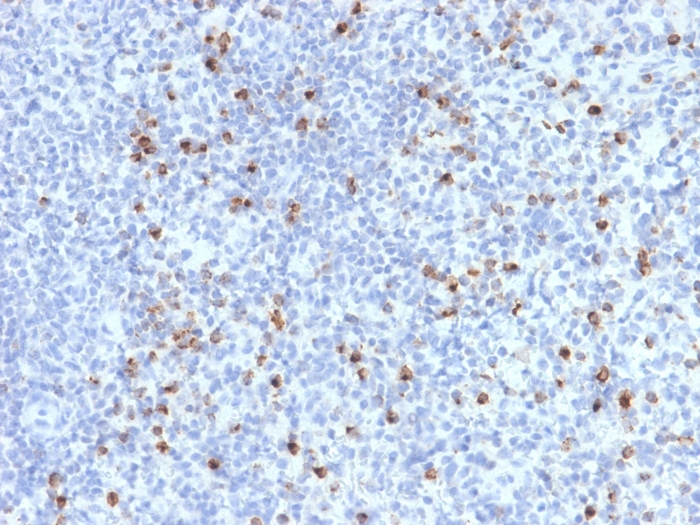
ApplicationsImmunoHistoChemistry, ImmunoHistoChemistry Paraffin
Product group Antibodies
ReactivityBovine, Human, Mouse
TargetFBN1
Overview
- SupplierBiotium
- Product NamePerforin-1 (Pore Forming Protein) (Apoptosis Marker)(PRF1/2470), CF405S conjugate, 0.1mg/mL
- Delivery Days Customer9
- ApplicationsImmunoHistoChemistry, ImmunoHistoChemistry Paraffin
- CertificationResearch Use Only
- ClonalityMonoclonal
- Clone IDPRF1/2470
- Concentration0.1 mg/ml
- ConjugateOther Conjugate
- Gene ID2200
- Target nameFBN1
- Target descriptionfibrillin 1
- Target synonymsACMICD; asprosin; ECTOL1; epididymis secretory sperm binding protein; FBN; fibrillin 15; fibrillin-1; fibrillin-1 preproprotein; GPHYSD2; MASS; MFLS; MFS1; OCTD; SGS; SSKS; WMS; WMS2
- HostMouse
- IsotypeIgG2c
- Protein IDP14222
- Protein NamePerforin-1
- Scientific DescriptionPerforin is a pore-forming protein that leads to osmotic lysis of the target cells and subsequently enables granzymes to enter the target cells and activate apoptosis. Perforin has structural and functional similarities to complement component 9 (C9). Like C9, this protein creates transmembrane tubules and is capable of lysing non-specifically a variety of target cells. It is one of the main cytolytic proteins of cytolytic granules, and is known to be a key effector molecule for T-cell- and natural killer-cell-mediated cytolysis. Defects in this gene cause familial hemophagocytic lymphohistiocytosis type 2 (HPLH2), a rare and lethal autosomal recessive disorder of early childhood. The expression of perforin is reportedly upregulated in activated CD8 T-cells, natural killer cells and some CD4 T-cells. Primary antibodies are available purified, or with a selection of fluorescent CF® Dyes and other labels. CF® Dyes offer exceptional brightness and photostability. Note: Conjugates of blue fluorescent dyes like CF®405S and CF®405M are not recommended for detecting low abundance targets, because blue dyes have lower fluorescence and can give higher non-specific background than other dye colors.
- SourceAnimal
- ReactivityBovine, Human, Mouse
- Storage Instruction2°C to 8°C
- UNSPSC12352203