

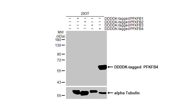
Non-transfected (–) and transfected (+) 293T whole cell extracts (30 μg) were separated by 10% SDS-PAGE, and the membrane was blotted with PFKFB4 antibody [HL2204] (GTX638208) diluted at 1:5000. The HRP-conjugated anti-rabbit IgG antibody (GTX213110-01) was used to detect the primary antibody.
![Various whole cell extracts (30 μg) were separated by 10% SDS-PAGE, and the membrane was blotted with PFKFB4 antibody [HL2204] (GTX638208) diluted at 1:1000. The HRP-conjugated anti-rabbit IgG antibody (GTX213110-01) was used to detect the primary antibody, and the signal was developed with Trident ECL plus-Enhanced. Corresponding RNA expression data for the same cell lines are based on Human Protein Atlas program.](https://www.genetex.com/upload/website/prouct_img/normal/GTX638208/GTX638208_T-44942_20230602_WB_TPM_watermark_23060622_403.webp "Various whole cell extracts (30 μg) were separated by 10% SDS-PAGE, and the membrane was blotted with PFKFB4 antibody [HL2204] (GTX638208) diluted at 1:1000. The HRP-conjugated anti-rabbit IgG antibody (GTX213110-01) was used to detect the primary antibody, and the signal was developed with Trident ECL plus-Enhanced. Corresponding RNA expression data for the same cell lines are based on Human Protein Atlas program.")
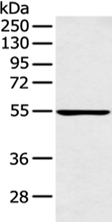
![Various whole cell extracts (30 μg) were separated by 10% SDS-PAGE, and the membrane was blotted with PFKFB4 antibody [HL2204] (GTX638208) diluted at 1:1000. The HRP-conjugated anti-rabbit IgG antibody (GTX213110-01) was used to detect the primary antibody.](https://www.genetex.com/upload/website/prouct_img/normal/GTX638208/GTX638208_45117_20230728_WB_23073119_424.webp "Various whole cell extracts (30 μg) were separated by 10% SDS-PAGE, and the membrane was blotted with PFKFB4 antibody [HL2204] (GTX638208) diluted at 1:1000. The HRP-conjugated anti-rabbit IgG antibody (GTX213110-01) was used to detect the primary antibody.")
![Various whole cell extracts (30 μg) were separated by 10% SDS-PAGE, and the membrane was blotted with PFKFB4 antibody [HL2204] (GTX638208) diluted at 1:1000. The HRP-conjugated anti-rabbit IgG antibody (GTX213110-01) was used to detect the primary antibody.](https://www.genetex.com/upload/website/prouct_img/normal/GTX638208/GTX638208_45117_20230818_WB_M_23082201_206.webp "Various whole cell extracts (30 μg) were separated by 10% SDS-PAGE, and the membrane was blotted with PFKFB4 antibody [HL2204] (GTX638208) diluted at 1:1000. The HRP-conjugated anti-rabbit IgG antibody (GTX213110-01) was used to detect the primary antibody.")
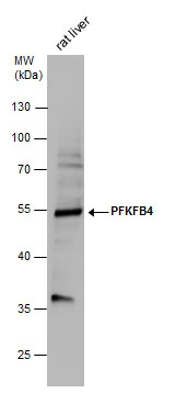
![Whole cell extract (30 μg) was separated by 10% SDS-PAGE, and the membrane was blotted with PFKFB4 antibody [HL2204] (GTX638208) diluted at 1:1000. The HRP-conjugated anti-rabbit IgG antibody (GTX213110-01) was used to detect the primary antibody.](https://www.genetex.com/upload/website/prouct_img/normal/GTX638208/GTX638208_45117_20230818_WB_R_23082201_574.webp "Whole cell extract (30 μg) was separated by 10% SDS-PAGE, and the membrane was blotted with PFKFB4 antibody [HL2204] (GTX638208) diluted at 1:1000. The HRP-conjugated anti-rabbit IgG antibody (GTX213110-01) was used to detect the primary antibody.")
![PFKFB4 antibody [HL2204] detects PFKFB4 protein at cell membrane and cytoplasm by immunohistochemical analysis. Sample: Paraffin-embedded mouse spleen. PFKFB4 stained by PFKFB4 antibody [HL2204] (GTX638208) diluted at 1:100. Antigen Retrieval: Citrate buffer, pH 6.0, 15 min](https://www.genetex.com/upload/website/prouct_img/normal/GTX638208/GTX638208_T-44942_20230816_IHC-P_M_23090619_908.webp "PFKFB4 antibody [HL2204] detects PFKFB4 protein at cell membrane and cytoplasm by immunohistochemical analysis. Sample: Paraffin-embedded mouse spleen. PFKFB4 stained by PFKFB4 antibody [HL2204] (GTX638208) diluted at 1:100. Antigen Retrieval: Citrate buffer, pH 6.0, 15 min")
![PFKFB4 antibody [HL2204] detects PFKFB4 protein at cell membrane and cytoplasm by immunohistochemical analysis. Sample: Paraffin-embedded rat spleen. PFKFB4 stained by PFKFB4 antibody [HL2204] (GTX638208) diluted at 1:100. Antigen Retrieval: Citrate buffer, pH 6.0, 15 min](https://www.genetex.com/upload/website/prouct_img/normal/GTX638208/GTX638208_T-44942_20230816_IHC-P_R_23090619_336.webp "PFKFB4 antibody [HL2204] detects PFKFB4 protein at cell membrane and cytoplasm by immunohistochemical analysis. Sample: Paraffin-embedded rat spleen. PFKFB4 stained by PFKFB4 antibody [HL2204] (GTX638208) diluted at 1:100. Antigen Retrieval: Citrate buffer, pH 6.0, 15 min")
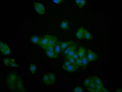
![PFKFB4 antibody [HL2204] detects PFKFB4 protein at cytoplasm and nucleus by immunofluorescent analysis. Sample: U87-MG cells were fixed in ice-cold MeOH for 5 min. Green: PFKFB4 stained by PFKFB4 antibody [HL2204] (GTX638208) diluted at 1:500. Red: alpha Tubulin, a cytoskeleton marker, stained by alpha Tubulin antibody [GT114] (GTX628802) diluted at 1:1000. Blue: Fluoroshield with DAPI (GTX30920).](https://www.genetex.com/upload/website/prouct_img/normal/GTX638208/GTX638208_45117_20240301_ICC_IF_24031823_953.webp "PFKFB4 antibody [HL2204] detects PFKFB4 protein at cytoplasm and nucleus by immunofluorescent analysis. Sample: U87-MG cells were fixed in ice-cold MeOH for 5 min. Green: PFKFB4 stained by PFKFB4 antibody [HL2204] (GTX638208) diluted at 1:500. Red: alpha Tubulin, a cytoskeleton marker, stained by alpha Tubulin antibody [GT114] (GTX628802) diluted at 1:1000. Blue: Fluoroshield with DAPI (GTX30920).")
Non-transfected (–) and transfected (+) 293T whole cell extracts (30 μg) were separated by 10% SDS-PAGE, and the membrane was blotted with PFKFB4 antibody [HL2204] (GTX638208) diluted at 1:5000. The HRP-conjugated anti-rabbit IgG antibody (GTX213110-01) was used to detect the primary antibody.
PFKFB4 antibody [HL2204]
GTX638208
ApplicationsImmunoFluorescence, Western Blot, ImmunoCytoChemistry, ImmunoHistoChemistry, ImmunoHistoChemistry Paraffin
Product group Antibodies
ReactivityHuman, Mouse, Rat
TargetPFKFB4
Overview
- SupplierGeneTex
- Product NamePFKFB4 antibody [HL2204]
- Delivery Days Customer9
- Application Supplier NoteWB: 1:1000-1:10000. *Optimal dilutions/concentrations should be determined by the researcher.Not tested in other applications.
- ApplicationsImmunoFluorescence, Western Blot, ImmunoCytoChemistry, ImmunoHistoChemistry, ImmunoHistoChemistry Paraffin
- CertificationResearch Use Only
- ClonalityMonoclonal
- Clone IDHL2204
- Concentration1 mg/ml
- ConjugateUnconjugated
- Gene ID5210
- Target namePFKFB4
- Target description6-phosphofructo-2-kinase/fructose-2,6-biphosphatase 4
- Target synonyms6-phosphofructo-2-kinase/fructose-2,6-bisphosphatase 4, 6PF-2-K/Fru-2,6-P2ase 4, 6PF-2-K/Fru-2,6-P2ase testis-type isozyme, PFK/FBPase 4, bifunctional enzyme with kinase and biphosphatase activities
- HostRabbit
- IsotypeIgG
- Protein IDQ16877
- Protein Name6-phosphofructo-2-kinase/fructose-2,6-bisphosphatase 4
- Scientific DescriptionThe protein encoded by this gene is one of four bifunctional kinase/phosphatases that regulate the concentration of the glycolytic byproduct fructose-2,6-bisphosphate (F2,6BP). The encoded protein is highly expressed in cancer cells and is induced by hypoxia. This protein is essential to the survival of cancer cells under conditions of hypoxia, because it increases the amount of F2,6BP and ATP at a time when the cell cannot produce much of them. This finding suggests that this protein may be a good target for disruption in cancer cells, hopefully imperiling their survival. Several transcript variants encoding different isoforms have been found for this gene. [provided by RefSeq, Nov 2015]
- ReactivityHuman, Mouse, Rat
- Storage Instruction-20°C or -80°C,2°C to 8°C
- UNSPSC41116161
Datasheet
Related products
Product group Antibodies
PFKFB4 AntibodyCSB-PA613704LA01HU
ApplicationsImmunoFluorescence, ELISA
ReactivityHuman
TargetPFKFB4
- SizePrice

Product group Antibodies
Anti-PFKFB4 Antibody Picoband(r)A07028-1-CARRIER-FREE
ApplicationsWestern Blot, ELISA
ReactivityHuman
TargetPFKFB4
- SizePrice

Product group Antibodies
Anti-PFKFB4 AntibodyHPA047719
ApplicationsImmunoCytoChemistry
ReactivityHuman
TargetPFKFB4
- SizePrice
Product group Antibodies

PFKFB4 AntibodyLS-C669650
ApplicationsImmunoFluorescence, ELISA
ReactivityHuman
TargetPFKFB4
- SizePrice
![Various whole cell extracts (30 μg) were separated by 10% SDS-PAGE, and the membrane was blotted with PFKFB4 antibody [HL3254] (GTX640901) diluted at 1:1000. The HRP-conjugated anti-rabbit IgG antibody (GTX213110-01) was used to detect the primary antibody. Corresponding RNA expression data for the same cell lines are based on Human Protein Atlas program.](https://www.genetex.com/upload/website/prouct_img/normal/GTX640901/GTX640901_T-45523_20240920_WB_TPM_watermark_24092600_654.webp)
Product group Antibodies
PFKFB4 antibody [HL3254]GTX640901
ApplicationsWestern Blot
ReactivityHuman, Rat
TargetPFKFB4
- SizePrice
![ICC/IF analysis of HepG2 cells using GTX83904 PFKFB4 antibody [1C8].](https://www.genetex.com/upload/website/prouct_img/normal/GTX83904/GTX83904_871_ICCIF_w_23061420_943.webp)
Product group Antibodies
PFKFB4 antibody [1C8]GTX83904
ApplicationsImmunoFluorescence, Western Blot, ImmunoCytoChemistry
ReactivityHuman
TargetPFKFB4
- SizePrice
Product group Antibodies
PFKFB4 antibodyGTX107755
ApplicationsImmunoFluorescence, Western Blot, ImmunoCytoChemistry, ImmunoHistoChemistry, ImmunoHistoChemistry Paraffin
ReactivityHuman, Mouse, Rat
TargetPFKFB4
- SizePrice