

PLK1 (Marker of Mitosis)(AZ44), CF594 conjugate, 0.1mg/mL [26628-22-8]
BNC943101
ApplicationsImmunoPrecipitation, Western Blot
Product group Antibodies
ReactivityXenopus
TargetDYzEms10
Overview
- SupplierBiotium
- Product NamePLK1 (Marker of Mitosis)(AZ44), CF594 conjugate, 0.1mg/mL [26628-22-8]
- Delivery Days Customer9
- ApplicationsImmunoPrecipitation, Western Blot
- CAS Number26628-22-8
- CertificationResearch Use Only
- ClonalityMonoclonal
- Clone IDAZ44
- Concentration0.1 mg/ml
- ConjugateOther Conjugate
- Gene ID28141
- Target nameDYzEms10
- Target descriptionDNA segment, Chr Y, repetitive sequence, Elizabeth M. Simpson 10
- HostMouse
- IsotypeIgG1
- Protein IDP70032
- Protein NameSerine/threonine-protein kinase PLK1
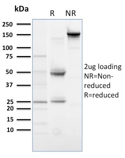
- Scientific DescriptionPlk (for polo-like kinase) encodes a serine/threonine kinase that is closely related to polo and CDC5, genes that are required for passage through mitosis in Drosophila and Saccharomyces, respectively. Polo and Cdc5 both code for proteins that are involved in regulating the function of the mitotic spindle. Plk protein accumulates in the cell during the S and G2 phases of the cell cycle; Plk protein content and catalytic activity peak at the onset of mitosis, followed by a rapid reduction after mitosis. Plk expression is detectable in mitotically active tissues such as colon and placenta, as well as in tumors of various origins. It has also been suggested that Plk may serve as a marker of cell proliferation. Required for recovery after DNA damage checkpoint and entry into mitosis. Required for kinetochore localization of BUB1B. Phosphorylates SGOL1. Required for spindle pole localization of isoform 3 of SGOL1 and plays a role in regulating its centriole cohesion function. Phosphorylates BORA, and thereby promotes the degradation of BORA. Contributes to the regulation of AURKA function. Regulates TP53 stability through phosphorylation of TOPORS. Useful for confirming that extracts have maintained CSF status and have not yet been activated into interphase. Primary antibodies are available purified, or with a selection of fluorescent CF® Dyes and other labels. CF® Dyes offer exceptional brightness and photostability. Note: Conjugates of blue fluorescent dyes like CF®405S and CF®405M are not recommended for detecting low abundance targets, because blue dyes have lower fluorescence and can give higher non-specific background than other dye colors.
- SourceAnimal
- ReactivityXenopus
- Storage Instruction2°C to 8°C,RT
- UNSPSC41116161