

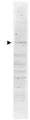
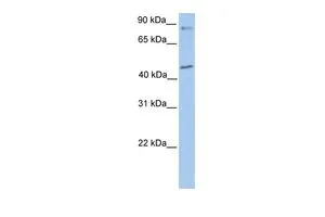
Western blot using GeneTex's Affinity Purified anti-POLK antibody shows detection of a band ~98 kDa corresponding to human POLK (arrowhead). Approximately 35 μg of a HeLa whole cell lysate was separated by 4-20% SDS-PAGE and transferred onto nitrocellulose. After blocking the membrane was probed with the primary antibody diluted to 1:1,000. Reaction occurred overnight at 4o followed by washes and reaction with a 1:20,000 dilution of IRDye800 conjugated rabbit anti-Goat IgG [H&L] MXHu for 45 min at room temperature. IRDye800 fluorescence image was captured using the OdysseyR Infrared Imaging System developed by LI-COR. IRDye is a trademark of LI-COR, Inc. Other detection systems will yield similar results.

Western blot using GeneTex's Affinity Purified anti-POLK antibody shows detection of a band ~98 kDa corresponding to human POLK (arrowhead). Approximately 35 μg of a HeLa whole cell lysate was separated by 4-20% SDS-PAGE and transferred onto nitrocellulose. After blocking the membrane was probed with the primary antibody diluted to 1:1,000. Reaction occurred overnight at 4o followed by washes and reaction with a 1:20,000 dilution of IRDye800 conjugated rabbit anti-Goat IgG [H&L] MXHu for 45 min at room temperature. IRDye800 fluorescence image was captured using the OdysseyR Infrared Imaging System developed by LI-COR. IRDye is a trademark of LI-COR, Inc. Other detection systems will yield similar results.
POLK antibody
GTX48495
ApplicationsWestern Blot, ELISA
Product group Antibodies
ReactivityHuman
TargetPOLK
Overview
- SupplierGeneTex
- Product NamePOLK antibody
- Delivery Days Customer9
- Application Supplier NoteWB: 1:500-1:3000. ELISA: 1:2000-1:10000. *Optimal dilutions/concentrations should be determined by the researcher.Not tested in other applications.
- ApplicationsWestern Blot, ELISA
- CertificationResearch Use Only
- ClonalityPolyclonal
- Concentration1.4 mg/ml
- ConjugateUnconjugated
- Gene ID51426
- Target namePOLK
- Target descriptionDNA polymerase kappa
- Target synonymsDINB1, DINP, POLQ, DNA polymerase kappa, polymerase (DNA directed) kappa
- HostGoat
- IsotypeIgG
- Protein IDQ9UBT6
- Protein NameDNA polymerase kappa
- Scientific DescriptionPOLK (also known as DNA polymerase kappa, DINB protein and DINP) is involved in DNA repair. POLK plays an important role in translesion synthesis, where the normal high-fidelity DNA polymerases cannot proceed and DNA synthesis stalls. Depending on the context, it inserts the correct base, but causes frequent base transitions, transversions and frameshifts. POLK lacks 3-5 proofreading exonuclease activity. POLK is a nuclear protein that binds to REV1L and PCNA. POLK is detected throughout the nucleus and at replication foci. Multiple alternative splice variants have been detected for this protein. POLK is found at low levels in testis, spleen, prostate and ovary and at very low levels in kidney, colon, brain, heart, liver, lung, placenta, pancreas and peripheral blood leukocytes.
- ReactivityHuman
- Storage Instruction-20°C or -80°C,2°C to 8°C
- UNSPSC41116161
Datasheet
Related products
Product group Antibodies
ApplicationsWestern Blot
ReactivityHuman, Mouse, Rat
TargetPOLK
- SizePrice
Product group Antibodies

POLK / DNA Polymerase Kappa AntibodyLS-C747206
ApplicationsWestern Blot
ReactivityHuman, Mouse, Rat
TargetPOLK
- SizePrice
Product group Antibodies
Anti-POLK AntibodyA39993
ApplicationsImmunoFluorescence, Western Blot
ReactivityHuman, Mouse, Rat
- SizePrice

Product group Antibodies
Anti-POLK AntibodyHPA057303
ApplicationsImmunoCytoChemistry
ReactivityHuman
TargetPOLK
- SizePrice
Product group Antibodies
POLK AntibodyCSB-PA887009LA01HU
ApplicationsWestern Blot, ELISA
ReactivityHuman, Mouse
TargetPOLK
- SizePrice
Product group Antibodies
Polk Polyclonal AntibodyCAC11361
ApplicationsWestern Blot, ELISA
ReactivityMouse
TargetPOLK
- SizePrice
Product group Antibodies
POLK antibody, InternalGTX46189
ApplicationsWestern Blot
ReactivityHuman
TargetPOLK
- SizePrice
Product group Antibodies
Anti-POLK Antibody144-12052
ApplicationsWestern Blot
ReactivityHuman, Mouse
TargetPOLK
- SizePrice
Product group Antibodies
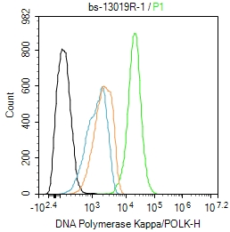
POLK Polyclonal AntibodyBS-13019R
ApplicationsFlow Cytometry
ReactivityHuman
TargetPOLK
- SizePrice