

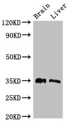
Western Blot Positive WB detected in: Mouse brain tissue, Rat liver tissue All lanes: PTF1A antibody at 2.7ug/ml Secondary Goat polyclonal to rabbit IgG at 1/50000 dilution Predicted band size: 35 kDa Observed band size: 35 kDa

")
Western Blot Positive WB detected in: Mouse brain tissue, Rat liver tissue All lanes: PTF1A antibody at 2.7ug/ml Secondary Goat polyclonal to rabbit IgG at 1/50000 dilution Predicted band size: 35 kDa Observed band size: 35 kDa
PTF1A Antibody
CSB-PA018967LA01HU
ApplicationsImmunoFluorescence, Western Blot, ELISA, ImmunoHistoChemistry
Product group Antibodies
ReactivityHuman, Mouse, Rat
TargetPTF1A
Overview
- SupplierCusabio
- Product NamePTF1A Antibody
- Delivery Days Customer20
- ApplicationsImmunoFluorescence, Western Blot, ELISA, ImmunoHistoChemistry
- CertificationResearch Use Only
- ClonalityPolyclonal
- ConjugateUnconjugated
- Gene ID256297
- Target namePTF1A
- Target descriptionpancreas associated transcription factor 1a
- Target synonymsPACA, PAGEN2, PTF1-p48, bHLHa29, p48, pancreas transcription factor 1 subunit alpha, bHLH transcription factor p48, class A basic helix-loop-helix protein 29, class II bHLH protein PTF1A, exocrine pancreas-specific transcription factor p48, p48 DNA-binding subunit of transcription factor PTF1, pancreas specific transcription factor, 1a
- HostRabbit
- IsotypeIgG
- Protein IDQ7RTS3
- Protein NamePancreas transcription factor 1 subunit alpha
- Scientific DescriptionTranscription factor implicated in the cell fate determination in various organs. Binds to the E-box consensus sequence 5-CANNTG-3. Plays a role in early and late pancreas development and differentiation. Important for determining whether cells allocated to the pancreatic buds continue towards pancreatic organogenesis or revert back to duodenal fates. May be involved in the maintenance of exocrine pancreas-specific gene expression including ELA1 and amylase. Required for the formation of pancreatic acinar and ductal cells. Plays an important role in cerebellar development. Directly regulated by FOXN4 and RORC during retinal development, FOXN4-PTF1A pathway plays a central role in directing the differentiation of retinal progenitors towards horizontal and amacrine fates.
- ReactivityHuman, Mouse, Rat
- Storage Instruction-20°C or -80°C
- UNSPSC41116161
Related products

Product group Antibodies
Anti-PTF1A AntibodyHPA073033
ApplicationsImmunoCytoChemistry
ReactivityHuman
TargetPTF1A
- SizePrice

Product group Antibodies
Anti-PTF1A Antibody Picoband(r)A03891-2-CARRIER-FREE
ApplicationsFlow Cytometry, ImmunoFluorescence, Western Blot, ELISA, ImmunoCytoChemistry
ReactivityHuman, Mouse, Rat
TargetPTF1A
- SizePrice
Product group Antibodies

PTF1A AntibodyLS-C679241
ApplicationsImmunoFluorescence, ELISA, ImmunoHistoChemistry, ImmunoHistoChemistry Paraffin
ReactivityHuman, Mouse, Rat
TargetPTF1A
- SizePrice
Product group Antibodies
Ptf1A Polyclonal AntibodyCAC08024
ApplicationsImmunoFluorescence, Western Blot, ELISA, ImmunoHistoChemistry
ReactivityMouse, Rat
TargetPTF1A
- SizePrice
Product group Antibodies
PTF1A AntibodyPACO51902
ApplicationsImmunoFluorescence, Western Blot, ELISA, ImmunoHistoChemistry
ReactivityHuman, Mouse, Rat
TargetPTF1A
- SizePrice
Product group Antibodies
PTF1A antibodyGTX129525
ApplicationsWestern Blot
ReactivityHuman, Mouse, Rat
TargetPTF1A
- SizePrice
Product group Antibodies
Anti-PTF1A Antibody101-11348
ApplicationsWestern Blot, ELISA
TargetPTF1A
- SizePrice