

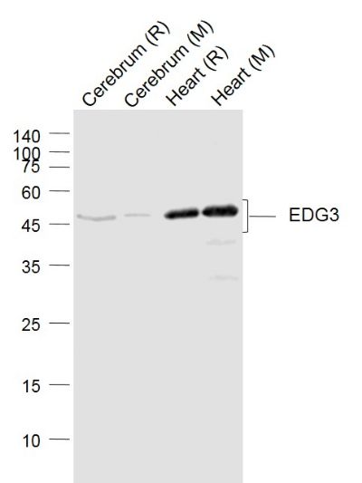
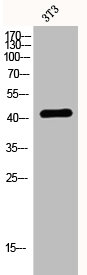
Rabbit anti Human Sphingosine 1 Phosphate 3 Receptor (EDG-3) CT
X1589P
ApplicationsImmunoPrecipitation, Western Blot
Product group Antibodies
ReactivityHuman, Mouse
TargetS1PR3
Product X1589P is not available
Product not available
There may be an alternative product available, please contact our technical support team.
Overview
- SupplierNordic-MUbio
- Product NameRabbit anti Human Sphingosine 1 Phosphate 3 Receptor (EDG-3) CT
- Delivery Days Customer7
- Application Supplier NoteAntibody can be used for Western blotting (5-10 microg/ml) and Immunoprecipitation (see reference 4). Optimal concentration should be evaluated by serial dilutions. Due to low expression of EDG receptors, we recommend use of Pierce Femto Signal substrate for western blot development.
- ApplicationsImmunoPrecipitation, Western Blot
- Applications SupplierWestern Blotting;Immunoprecipitation
- CertificationResearch Use Only
- ClonalityPolyclonal
- ConjugateUnconjugated
- Gene ID1903
- Target nameS1PR3
- Target descriptionsphingosine-1-phosphate receptor 3
- Target synonymsC9orf108, C9orf47, EDG-3, EDG3, LPB3, S1P3, bA791O21.3, sphingosine 1-phosphate receptor 3, G protein-coupled receptor, endothelial differentiation gene-3, S1P receptor 3, S1P receptor EDG3, S1P receptor Edg-3, endothelial differentiation G-protein coupled receptor 3, endothelial differentiation, sphingolipid G-protein-coupled receptor, 3, sphingosine 1-phosphate receptor Edg-3, uncharacterized protein C9orf47
- HostRabbit
- Protein IDQ99500
- Protein NameSphingosine 1-phosphate receptor 3
- Scientific DescriptionEndothelial cell differentiation gene 3 C-terminal; Sphingosine-1-Phosphate Receptor-3 (S1P3)
- Shelf life instructionSee expiration date on vial
- SourceRabbits were immunized with a synthetic peptide derived from the C terminal of the EDG3 (S1P3) proteins
- ReactivityHuman, Mouse
- Reactivity SupplierHuman;Mouse
- UNSPSC12352203
Related products
Product group Antibodies
Anti-EDG3 (N-term) Antibody102-26956
ApplicationsWestern Blot
TargetS1PR3
- SizePrice

Product group Antibodies
Anti-EDG3 AntibodyA100005
ApplicationsImmunoFluorescence, Western Blot, ELISA
ReactivityHuman
- SizePrice
Product group Antibodies
References
EDG3 Polyclonal AntibodyBS-7541R
ApplicationsFlow Cytometry, ImmunoFluorescence, Western Blot, ELISA, ImmunoCytoChemistry, ImmunoHistoChemistry, ImmunoHistoChemistry Frozen, ImmunoHistoChemistry Paraffin
ReactivityBovine, Human, Mouse, Rabbit, Rat
TargetS1PR3
- SizePrice
Product group Antibodies
S1PR3 AntibodyCSB-PA002248
ApplicationsImmunoFluorescence, Western Blot, ELISA
ReactivityHuman
TargetS1PR3
- SizePrice
Product group Antibodies

Goat anti-EDG3EB09356
ApplicationsELISA, ImmunoHistoChemistry
ReactivityHuman
TargetS1PR3
- SizePrice

Product group Antibodies
References
ApplicationsImmunoFluorescence, Western Blot, ImmunoCytoChemistry, ImmunoHistoChemistry
ReactivityHuman
TargetS1PR3
- SizePrice
Product group Antibodies

SIPR3 / EDG3 / S1P3 AntibodyLS-C402838
ApplicationsWestern Blot, ELISA
ReactivityHuman, Mouse
TargetS1PR3
- SizePrice
Product group Antibodies
SIPR3 antibodyGTX77925
ApplicationsImmunoHistoChemistry, ImmunoHistoChemistry Paraffin
ReactivityHuman
TargetS1PR3
- SizePrice
Product group Antibodies
Anti-PHKG2 AntibodyCAB14040
ApplicationsWestern Blot, ELISA
ReactivityHuman
TargetS1PR3
- SizePrice