

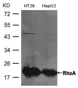
Western blot analysis of extracts from HT29 and HepG2 cells using RhoA Antibody.
Western blot analysis of extracts from HT29 and HepG2 cells using RhoA Antibody.
RHOA Antibody
CSB-PA042169
ApplicationsWestern Blot, ELISA
Product group Antibodies
ReactivityHuman, Mouse, Rat
TargetRHOA
Overview
- SupplierCusabio
- Product NameRHOA Antibody
- Delivery Days Customer20
- ApplicationsWestern Blot, ELISA
- CertificationResearch Use Only
- ClonalityPolyclonal
- ConjugateUnconjugated
- Gene ID387
- Target nameRHOA
- Target descriptionras homolog family member A
- Target synonymsAplysia ras-related homolog 12; ARH12; ARHA; EDFAOB; epididymis secretory sperm binding protein; oncogene RHO H12; RHO12; RHOH12; small GTP binding protein RhoA; transforming protein RhoA
- HostRabbit
- IsotypeIgG
- Protein IDP61586
- Protein NameTransforming protein RhoA
- Scientific DescriptionRegulates a signal transduction pathway linking plasma membrane receptors to the assembly of focal adhesions and actin stress fibers. Involved in a microtubule-dependent signal that is required for the myosin contractile ring formation during cell cycle cytokinesis. Plays an essential role in cleavage furrow formation. Required for the apical junction formation of keratinocyte cell-cell adhesion. Serves as a target for the yopT cysteine peptidase from Yersinia pestis, vector of the plague, and Yersinia pseudotuberculosis, which causes gastrointestinal disorders. Stimulates PKN2 kinase activity. May be an activator of PLCE1. Activated by ARHGEF2, which promotes the exchange of GDP for GTP. Essential for the SPATA13-mediated regulation of cell migration and adhesion assembly and disassembly. The MEMO1-RHOA-DIAPH1 signaling pathway plays an important role in ERBB2-dependent stabilization of microtubules at the cell cortex. It controls the localization of APC and CLASP2 to the cell membrane, via the regulation of GSK3B activity. In turn, membrane-bound APC allows the localization of the MACF1 to the cell membrane, which is required for microtubule capture and stabilization.
- ReactivityHuman, Mouse, Rat
- Storage Instruction-20°C or -80°C
- UNSPSC12352203