

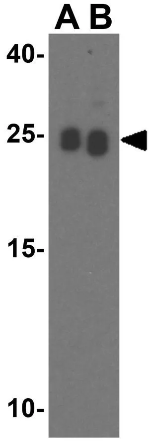
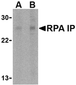
WB analysis of Jurkat cell lysate using GTX85485 RPA Interacting Protein antibody. Working concentration : (A) 0.5 and (B) 1 μg/ml
WB analysis of Jurkat cell lysate using GTX85485 RPA Interacting Protein antibody. Working concentration : (A) 0.5 and (B) 1 μg/ml
RPA Interacting Protein antibody
GTX85485
ApplicationsWestern Blot, ELISA
Product group Antibodies
ReactivityHuman, Mouse, Rat
TargetRPAIN
Overview
- SupplierGeneTex
- Product NameRPA Interacting Protein antibody
- Delivery Days Customer9
- Application Supplier NoteWB: 0.5 - 1 microg/mL. *Optimal dilutions/concentrations should be determined by the researcher.Not tested in other applications.
- ApplicationsWestern Blot, ELISA
- CertificationResearch Use Only
- ClonalityPolyclonal
- Concentration1 mg/ml
- ConjugateUnconjugated
- Gene ID84268
- Target nameRPAIN
- Target descriptionRPA interacting protein
- Target synonymsHRIP, RIP, RPA-interacting protein, RAP interaction protein, nuclear transporter
- HostRabbit
- IsotypeIgG
- Protein IDQ86UA6
- Protein NameRPA-interacting protein
- Scientific DescriptionReplication protein A (RPA) is a single-stranded-DNA binding protein involved in numerous eukaryotic DNA processes including replication, repair and recombination. RPA interacting protein (RPA IP) has been identified as an adapter protein that is involved in RPA nuclear import instead of the prototypical importin proteins that normally mediate nuclear import (2). Multiple isoforms of RPA IP are known to exist, with the longest isoform localized to the cytoplasm. Isoform 2 is sumoylated and is located in the PML nuclear body within the nucleus. It has been suggested that this isoform mediates the localization of the RPA complex into the PML nuclear body, thereby participating in RPA function in DNA metabolism.
- ReactivityHuman, Mouse, Rat
- Storage Instruction-20°C or -80°C,2°C to 8°C
- UNSPSC12352203
Datasheet
Related products
Product group Antibodies
Anti-RPAIN (N-term) Antibody102-22389
ApplicationsWestern Blot
TargetRPAIN
- SizePrice
Product group Antibodies
RPAIN AntibodyCSB-PA020093GA01HU
ApplicationsWestern Blot, ELISA, ImmunoHistoChemistry
ReactivityHuman
TargetRPAIN
- SizePrice
Product group Antibodies
ApplicationsWestern Blot, ELISA, ImmunoHistoChemistry
ReactivityHuman, Mouse, Rat
- SizePrice
Product group Antibodies

IHC-plus(tm) RPAIN AntibodyLS-B1396
ApplicationsWestern Blot, ELISA, ImmunoHistoChemistry, ImmunoHistoChemistry Paraffin
ReactivityHuman, Mouse, Rat
TargetRPAIN
- SizePrice

Product group Antibodies
Anti-RPAIN AntibodyHPA023924
ApplicationsImmunoHistoChemistry
ReactivityHuman
TargetRPAIN
- SizePrice
Product group Antibodies
RPA Interacting Protein antibodyGTX85486
ApplicationsWestern Blot, ELISA, ImmunoHistoChemistry, ImmunoHistoChemistry Paraffin
ReactivityHuman, Mouse, Rat
TargetRPAIN
- SizePrice

Product group Antibodies
Anti-RPAIN Antibody Picoband(r)A11439-1-CARRIER-FREE
ApplicationsFlow Cytometry, Western Blot, ELISA
ReactivityHuman, Mouse
TargetRPAIN
- SizePrice