

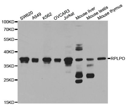
Western Blot analysis of NIH/3T3, A431 and Jurkat cell using RPLP0 Polyclonal Antibody at dilution of 1:750
Western Blot analysis of NIH/3T3, A431 and Jurkat cell using RPLP0 Polyclonal Antibody at dilution of 1:750
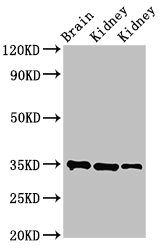
RPLP0 Polyclonal Antibody
E-AB-10583
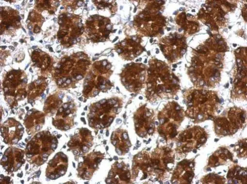
ApplicationsWestern Blot, ImmunoHistoChemistry
Product group Antibodies
TargetRPLP0
Overview
- SupplierElabscience
- Product NameRPLP0 Polyclonal Antibody
- Delivery Days Customer12
- ApplicationsWestern Blot, ImmunoHistoChemistry
- Applications SupplierELISA WB IHC
- CertificationResearch Use Only
- ClonalityPolyclonal
- Concentration0.4 mg/ml
- ConjugateUnconjugated
- Gene ID6175
- Target nameRPLP0
- Target descriptionribosomal protein lateral stalk subunit P0
- Target synonymsL10E, LP0, P0, PRLP0, RPP0, uL10, large ribosomal subunit protein uL10, 60S acidic ribosomal protein P0, 60S ribosomal protein L10E, acidic ribosomal phosphoprotein P0, neutral ribosomal phosphoprotein P0, ribosomal protein, large, P0
- HostRabbit
- IsotypeIgG
- Protein IDP05388
- Protein NameLarge ribosomal subunit protein uL10
- Scientific DescriptionRibosomes, the organelles that catalyze protein synthesis, consist of a small 40S subunit and a large 60S subunit. Together these subunits are composed of 4 RNA species and approximately 80 structurally distinct proteins. This gene encodes a ribosomal protein that is a component of the 60S subunit. The protein, which is the functional equivalent of the E. coli L10 ribosomal protein, belongs to the L10P family of ribosomal proteins. It is a neutral phosphoprotein with a C-terminal end that is nearly identical to the C-terminal ends of the acidic ribosomal phosphoproteins P1 and P2. The P0 protein can interact with P1 and P2 to form a pentameric complex consisting of P1 and P2 dimers, and a P0 monomer. The protein is located in the cytoplasm. Transcript variants derived from alternative splicing exist; they encode the same protein. As is typical for genes encoding ribosomal proteins, there are multiple processed pseudogenes of this gene dispersed through the genome.
- Storage Instruction-20°C
- UNSPSC41116161
MSDS
Related products
Product group Antibodies
RPLP0 AntibodyCSB-PA020336LA01HU
ApplicationsImmunoFluorescence, Western Blot, ELISA, ImmunoHistoChemistry
ReactivityHuman, Mouse, Rat
TargetRPLP0
- SizePrice
Product group Antibodies
Anti-RPLP0 AntibodyA30818
ApplicationsImmunoFluorescence, Western Blot, ImmunoHistoChemistry
ReactivityHuman, Mouse, Rat
- SizePrice

Product group Antibodies
Anti-RPLP0 AntibodyHPA003512
ApplicationsImmunoCytoChemistry, ImmunoHistoChemistry
ReactivityHuman
TargetRPLP0
- SizePrice

Product group Antibodies
Anti-RPLP0 Antibody Picoband(r)A04349-1-CARRIER-FREE
ApplicationsFlow Cytometry, ImmunoFluorescence, Western Blot, ELISA, ImmunoCytoChemistry
ReactivityHuman
TargetRPLP0
- SizePrice
Product group Antibodies

RPLP0 AntibodyLS-C401015
ApplicationsWestern Blot, ELISA, ImmunoHistoChemistry
ReactivityHuman, Mouse, Rat
TargetRPLP0
- SizePrice
Product group Antibodies
RPLP0 Polyclonal AntibodyCAC13912
ApplicationsImmunoFluorescence, Western Blot, ELISA, ImmunoHistoChemistry
ReactivityMouse, Rat
TargetRPLP0
- SizePrice
Product group Antibodies
RPLP0 antibodyGTX114730
ApplicationsWestern Blot, ImmunoHistoChemistry, ImmunoHistoChemistry Paraffin
ReactivityHuman
TargetRPLP0
- SizePrice
Product group Antibodies
Anti-RPLP0 Antibody144-05557
ApplicationsImmunoFluorescence, Western Blot, ImmunoHistoChemistry
ReactivityHuman, Mouse, Rat
TargetRPLP0
- SizePrice