

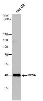
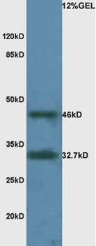
Western Blot analysis of A549, NIH/3T3 and 293T cell, Human hepatocellular carcinoma tissue and hela cell using RPSA Polyclonal Antibody at dilution of 1:425
Western Blot analysis of A549, NIH/3T3 and 293T cell, Human hepatocellular carcinoma tissue and hela cell using RPSA Polyclonal Antibody at dilution of 1:425
RPSA Polyclonal Antibody
E-AB-10457
ApplicationsWestern Blot
Product group Antibodies
TargetRPSA
Overview
- SupplierElabscience
- Product NameRPSA Polyclonal Antibody
- Delivery Days Customer12
- ApplicationsWestern Blot
- Applications SupplierELISA WB
- CertificationResearch Use Only
- ClonalityPolyclonal
- Concentration0.3 mg/ml
- ConjugateUnconjugated
- Gene ID3921
- Target nameRPSA
- Target descriptionribosomal protein SA
- Target synonyms37LRP, 67LR, ICAS, LAMBR, LAMR1, LBP, LBP/p40, LRP, LRP/LR, NEM/1CHD4, SA, lamR, p40, uS2, small ribosomal subunit protein uS2, 37 kDa laminin receptor, 37/67 kDa laminin receptor, 40S ribosomal protein SA, 67 kDa laminin receptor, colon carcinoma laminin-binding protein, laminin receptor 1 (67kD, ribosomal protein SA), laminin-binding protein precursor p40, multidrug resistance-associated protein MGr1-Ag
- HostRabbit
- IsotypeIgG
- Protein IDP08865
- Protein NameSmall ribosomal subunit protein uS2
- Scientific DescriptionLaminins, a family of extracellular matrix glycoproteins, are the major noncollagenous constituent of basement membranes. They have been implicated in a wide variety of biological processes including cell adhesion, differentiation, migration, signaling, neurite outgrowth and metastasis. Many of the effects of laminin are mediated through interactions with cell surface receptors. These receptors include members of the integrin family, as well as non-integrin laminin-binding proteins. This gene encodes a high-affinity, non-integrin family, laminin receptor 1. This receptor has been variously called 67 kD laminin receptor, 37 kD laminin receptor precursor (37LRP) and p40 ribosome-associated protein. The amino acid sequence of laminin receptor 1 is highly conserved through evolution, suggesting a key biological function. It has been observed that the level of the laminin receptor transcript is higher in colon carcinoma tissue and lung cancer cell line than their normal counterparts. Also, there is a correlation between the upregulation of this polypeptide in cancer cells and their invasive and metastatic phenotype. Multiple copies of this gene exist, however, most of them are pseudogenes thought to have arisen from retropositional events. Two alternatively spliced transcript variants encoding the same protein have been found for this gene
- Storage Instruction-20°C
- UNSPSC41116161
MSDS
Related products
Product group Antibodies
ReactivityHuman
TargetRPSA
- SizePrice
Product group Antibodies
TargetRPSA
- SizePrice
Product group Antibodies
RPSA AntibodyCSB-PA009782
ApplicationsWestern Blot, ELISA, ImmunoHistoChemistry
ReactivityHuman, Mouse, Rat
TargetRPSA
- SizePrice
Product group Antibodies

RPSA / Laminin Receptor AntibodyLS-C400882
ApplicationsWestern Blot, ELISA
ReactivityHuman, Mouse, Rat
TargetRPSA
- SizePrice
Product group Antibodies
RPSA Polyclonal AntibodyCAC13914
ApplicationsWestern Blot, ELISA
ReactivityMouse
TargetRPSA
- SizePrice

Product group Antibodies
Anti-67kDa Laminin Receptor/RPSA Antibody Picoband(r)PB10094-CARRIER-FREE
ApplicationsFlow Cytometry, ImmunoFluorescence, Western Blot, ImmunoCytoChemistry, ImmunoHistoChemistry, ImmunoHistoChemistry Frozen
ReactivityBovine, Canine, Chicken, Equine, Hamster, Human, Monkey
TargetRPSA
- SizePrice
Product group Antibodies
RPSA antibody [N1C3]GTX100831
ApplicationsWestern Blot, ImmunoHistoChemistry, ImmunoHistoChemistry Paraffin
ReactivityHuman, Mouse
TargetRPSA
- SizePrice
Product group Antibodies
References
RPSA Polyclonal AntibodyBS-0900R
ApplicationsFlow Cytometry, ImmunoFluorescence, Western Blot, ELISA, ImmunoCytoChemistry, ImmunoHistoChemistry, ImmunoHistoChemistry Frozen, ImmunoHistoChemistry Paraffin
ReactivityCanine, Human, Mouse, Rat
TargetRPSA
- SizePrice
Product group Antibodies
Anti-Mouse RPSA Antibody144-09008
ApplicationsWestern Blot
ReactivityHuman, Mouse
TargetRPSA
- SizePrice