

Search results: AMPK alpha 1
Product group Antibodies
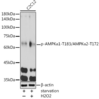
ApplicationsWestern Blot, ImmunoHistoChemistry
ReactivityHuman, Mouse, Rat
TargetPRKAA1
- SizePrice

Product group Antibodies
ApplicationsImmunoFluorescence, ImmunoPrecipitation, Western Blot, ImmunoCytoChemistry
ReactivityHuman
TargetPRKAA1
- SizePrice
Product group Antibodies
ApplicationsImmunoPrecipitation, Western Blot, ELISA, ImmunoHistoChemistry, ImmunoHistoChemistry Paraffin
- SizePrice
Product group Antibodies
AMPK alpha 2 Polyclonal Antibody, APC-Cy7 ConjugatedBS-2771R-APC-CY7
ApplicationsFlow Cytometry, ImmunoFluorescence, Western Blot, ImmunoCytoChemistry, ImmunoHistoChemistry, ImmunoHistoChemistry Frozen, ImmunoHistoChemistry Paraffin
ReactivityBovine, Canine, Human, Mouse, Porcine, Rabbit, Rat, Sheep
TargetPRKAA2
- SizePrice
Product group Antibodies
AMPK alpha 2 Polyclonal Antibody, PE-Cy5 ConjugatedBS-2771R-PE-CY5-TR
ApplicationsFlow Cytometry, Western Blot
TargetPRKAA2
- SizePrice
Product group Antibodies
AMPK alpha 2 Polyclonal Antibody, PE-Cy5 Conjugatedbs-2771R-PE-Cy5
ApplicationsFlow Cytometry, Western Blot
ReactivityBovine, Canine, Human, Mouse, Porcine, Rabbit, Rat, Sheep
TargetPRKAA2
- SizePrice
Product group Antibodies
AMPK alpha 2 Polyclonal Antibody, PE-Cy7 ConjugatedBS-2771R-PE-CY7-TR
ApplicationsFlow Cytometry, Western Blot
TargetPRKAA2
- SizePrice
Product group Antibodies
AMPK alpha 2 Polyclonal Antibody, PE-Cy7 Conjugatedbs-2771R-PE-Cy7
ApplicationsFlow Cytometry, Western Blot
ReactivityBovine, Canine, Human, Mouse, Porcine, Rabbit, Rat, Sheep
TargetPRKAA2
- SizePrice
Product group Antibodies
AMPK alpha 2 Polyclonal Antibody, PerCP ConjugatedBS-2771R-PERCP-TR
ApplicationsFlow Cytometry, ImmunoFluorescence, Western Blot, ImmunoCytoChemistry, ImmunoHistoChemistry, ImmunoHistoChemistry Frozen, ImmunoHistoChemistry Paraffin
TargetPRKAA2
- SizePrice
Product group Antibodies
AMPK alpha 2 Polyclonal Antibody, PerCP ConjugatedBS-2771R-PERCP
ApplicationsFlow Cytometry, ImmunoFluorescence, Western Blot, ImmunoCytoChemistry, ImmunoHistoChemistry, ImmunoHistoChemistry Frozen, ImmunoHistoChemistry Paraffin
ReactivityBovine, Canine, Human, Mouse, Porcine, Rabbit, Rat, Sheep
TargetPRKAA2
- SizePrice
Product group Antibodies
AMPK alpha 2 Polyclonal Antibody, PerCP ConjugatedBS-22269R-PERCP
ApplicationsImmunoFluorescence
ReactivityBovine, Canine, Equine, Human, Porcine, Rat, Sheep
TargetPRKAA2
- SizePrice
Product group Antibodies
ApplicationsFlow Cytometry, ImmunoFluorescence, Western Blot, ELISA, ImmunoCytoChemistry, ImmunoHistoChemistry, ImmunoHistoChemistry Frozen, ImmunoHistoChemistry Paraffin
ReactivityMouse, Rat
TargetPRKAA1
- SizePrice
Didn't find what you were looking for?
Search through our product groups to find the right product
Back to overview