Bio-Connect

Search results: B7H4

37 to 48 of 417 products
Sort by
Figure 1. Western blot analysis of VTCN1 using anti-VTCN1 antibody (A02821-3). Electrophoresis was performed on a 5-20% SDS-PAGE gel at 70V (Stacking gel) / 90V (Resolving gel) for 2-3 hours. The sample well of each lane was loaded with 30 ug of sample under reducing conditions. Lane 1: human MCF-7 whole cell lysates, Lane 2: human Hacat whole cell lysates, Lane 3: human A431 whole cell lysates, Lane 4: rat liver tissue lysates, Lane 5: rat RH35 whole cell lysates, Lane 6: mouse liver tissue lysates, Lane 7: moue liver tissue lysates, Lane 8: mouse HEPA1-6 whole cell lysates, After electrophoresis, proteins were transferred to a nitrocellulose membrane at 150 mA for 50-90 minutes. Blocked the membrane with 5% non-fat milk/TBS for 1.5 hour at RT. The membrane was incubated with rabbit anti-VTCN1 antigen affinity purified polyclonal antibody (Catalog # A02821-3) at 0.5 microg/mL overnight at 4°C, then washed with TBS-0.1%Tween 3 times with 5 minutes each and probed with a goat anti-rabbit IgG-HRP secondary antibody at a dilution of 1:5000 for 1.5 hour at RT. The signal is developed using an Enhanced Chemiluminescent detection (ECL) kit (Catalog # EK1002) with Tanon 5200 system. A specific band was detected for VTCN1 at approximately 75 kDa. The expected band size for VTCN1 is at 31 kDa.
Product group Antibodies
Boster Bio
ApplicationsFlow Cytometry, ImmunoFluorescence, Western Blot, ELISA, ImmunoCytoChemistry, ImmunoHistoChemistry
ReactivityHuman, Mouse, Rat
TargetVTCN1
  • SizePrice
Product group Antibodies
Bioss Antibodies
ApplicationsFlow Cytometry
ReactivityCanine, Human, Mouse, Rat
TargetVtcn1
  • SizePrice
Product group Antibodies
Bioss Antibodies
ApplicationsFlow Cytometry
ReactivityCanine, Human, Mouse, Rat
TargetVtcn1
  • SizePrice
Recombinant Human B7H4 Protein (Fc Tag)
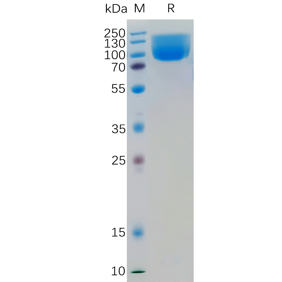
Product group Proteins / Signaling Molecules
Antibodies.com
ApplicationsELISA, Other Application
  • SizePrice