

Search results: IL22
Product group Antibodies
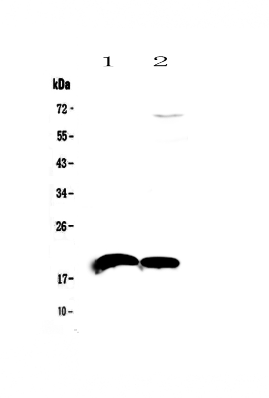
Anti-IL22 Antibody Picoband(r)A00963-3-APC
ApplicationsWestern Blot, ELISA
ReactivityMouse, Rat
TargetIl22
- SizePrice
Product group Antibodies
Anti-IL22 Antibody Picoband(r)A00963-3-BIOTIN
ApplicationsWestern Blot, ELISA
ReactivityMouse, Rat
TargetIl22
- SizePrice
Product group Antibodies
Anti-IL22 Antibody Picoband(r)A00963-3-CARRIER-FREE
ApplicationsWestern Blot, ELISA
ReactivityMouse, Rat
TargetIl22
- SizePrice
Product group Antibodies
Anti-IL22 Antibody Picoband(r)A00963-3-CY3
ApplicationsWestern Blot, ELISA
ReactivityMouse, Rat
TargetIl22
- SizePrice
Product group Antibodies
Anti-IL22 Antibody Picoband(r)A00963-3-DYLIGHT488
ApplicationsWestern Blot, ELISA
ReactivityMouse, Rat
TargetIl22
- SizePrice
Product group Antibodies
Anti-IL22 Antibody Picoband(r)A00963-3-DYLIGHT550
ApplicationsWestern Blot, ELISA
ReactivityMouse, Rat
TargetIl22
- SizePrice
Product group Antibodies
Anti-IL22 Antibody Picoband(r)A00963-3-DYLIGHT594
ApplicationsWestern Blot, ELISA
ReactivityMouse, Rat
TargetIl22
- SizePrice
Product group Antibodies
Anti-IL22 Antibody Picoband(r)A00963-3-FITC
ApplicationsWestern Blot, ELISA
ReactivityMouse, Rat
TargetIl22
- SizePrice
Product group Antibodies
Anti-IL22 Antibody Picoband(r)A00963-3-HRP
ApplicationsWestern Blot, ELISA
ReactivityMouse, Rat
TargetIl22
- SizePrice
Product group Antibodies
Anti-IL22 Antibody Picoband(r)A00963-3-IFLUOR647
ApplicationsWestern Blot, ELISA
ReactivityMouse, Rat
TargetIl22
- SizePrice
Product group Antibodies
Anti-IL22 Antibody Picoband(r)A00963-3-PE
ApplicationsWestern Blot, ELISA
ReactivityMouse, Rat
TargetIl22
- SizePrice
Product group Antibodies
Anti-IL22 Antibody Picoband(r)A00963-3
ApplicationsWestern Blot, ELISA
ReactivityMouse, Rat
TargetIl22
- SizePrice
Didn't find what you were looking for?
Search through our product groups to find the right product
Back to overview