

Search results: TAF12
Product group Antibodies
Anti-TAF12 AntibodyA30966
ApplicationsWestern Blot, ImmunoHistoChemistry
ReactivityHuman, Mouse, Rat
- SizePrice
Product group Antibodies
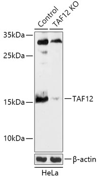
Anti-TAF12 AntibodyA92845
ApplicationsWestern Blot
ReactivityHuman, Mouse, Rat
- SizePrice
Product group Antibodies
Anti-TAF12 Antibody144-05421
ApplicationsWestern Blot
ReactivityHuman, Mouse, Rat
TargetTAF12
- SizePrice
Product group Antibodies
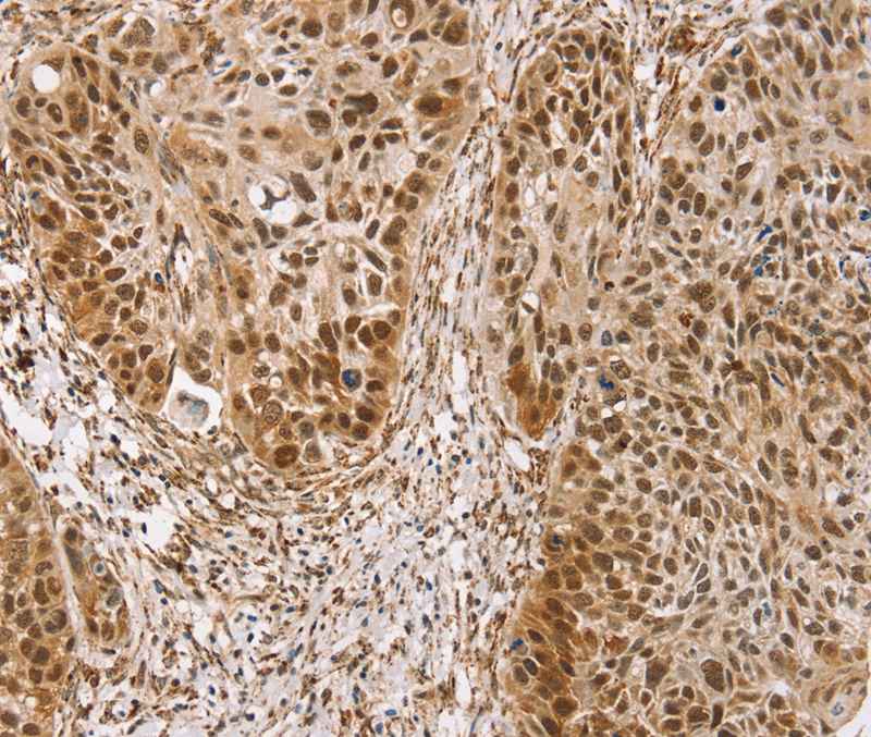
Anti-TAF12 AntibodyA46319
ApplicationsImmunoHistoChemistry
ReactivityHuman
- SizePrice
Product group Antibodies
TAF12 Polyclonal AntibodyBS-5142R
ApplicationsImmunoFluorescence, ELISA, ImmunoCytoChemistry, ImmunoHistoChemistry, ImmunoHistoChemistry Frozen, ImmunoHistoChemistry Paraffin
ReactivityBovine, Canine, Chicken, Human, Mouse, Porcine, Rat
TargetTAF12
- SizePrice

Product group Antibodies
Anti-TAF12 AntibodyA06944-1
ApplicationsWestern Blot, ImmunoHistoChemistry
ReactivityHuman, Mouse, Rat
TargetTAF12
- SizePrice
Product group Antibodies
TAF12 Antibody, HRP conjugatedCSB-PA624102LB01HU
ApplicationsELISA
ReactivityHuman
TargetTAF12
- SizePrice
Product group Antibodies
TAF12 Antibody, FITC conjugatedCSB-PA624102LC01HU
ReactivityHuman
TargetTAF12
- SizePrice
Product group Antibodies
TAF12 Antibody, Biotin conjugatedCSB-PA624102LD01HU
ApplicationsELISA
ReactivityHuman
TargetTAF12
- SizePrice
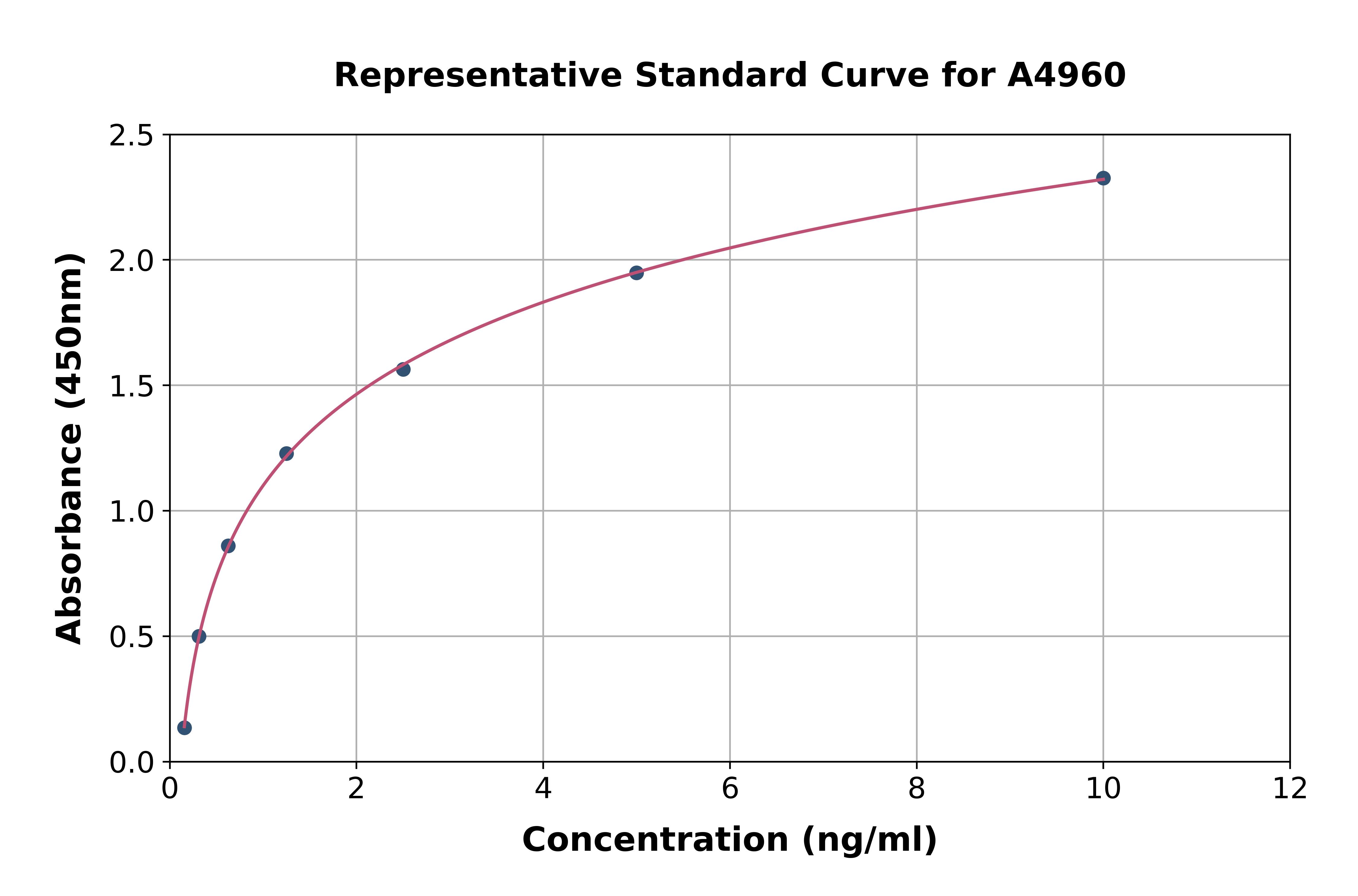
Product group Assays
Assay Sample TypeTissue homogenates and other biological fluids.
ReactivityMouse
- SizePrice
Product group Antibodies
Anti-TAF12 Antibody Picoband(r)A06944-1-BIOTIN
ApplicationsWestern Blot, ImmunoHistoChemistry
ReactivityHuman, Mouse, Rat
TargetTAF12
- SizePrice
Didn't find what you were looking for?
Search through our product groups to find the right product
Back to overview