

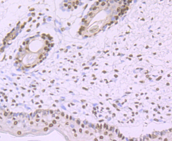
IHC-P analysis of human skin tissue section using GTX01165 SF3B1 antibody [JB40-32].
![IHC-P analysis of rat brain tissue section using GTX01165 SF3B1 antibody [JB40-32].](https://www.genetex.com/upload/website/prouct_img/normal/GTX01165/GTX01165_20200303_IHC-P_649_w_23053121_312.webp "IHC-P analysis of rat brain tissue section using GTX01165 SF3B1 antibody [JB40-32].")
![IHC-P analysis of human kidney tissue section using GTX01165 SF3B1 antibody [JB40-32].](https://www.genetex.com/upload/website/prouct_img/normal/GTX01165/GTX01165_20200303_IHC-P_652_w_23053121_301.webp "IHC-P analysis of human kidney tissue section using GTX01165 SF3B1 antibody [JB40-32].")
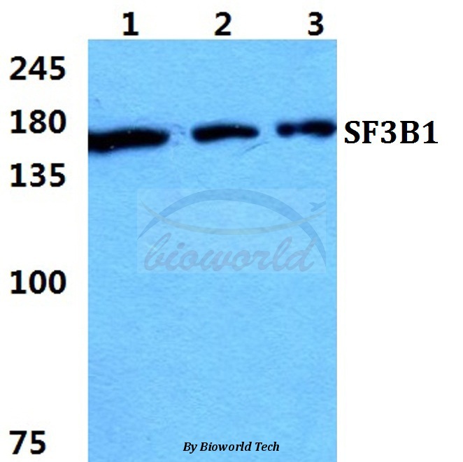
![Various whole cell extracts (30 μg) were separated by 5% SDS-PAGE, and the membrane was blotted with SF3B1 antibody [JB40-32] (GTX01165) diluted at 1:500. The HRP-conjugated anti-rabbit IgG antibody (GTX213110-01) was used to detect the primary antibody. Corresponding RNA expression data for the same cell lines are based on Human Protein Atlas program.](https://www.genetex.com/upload/website/prouct_img/normal/GTX01165/GTX01165_HL0222_20200228_WB_TPM_watermark_w_23053121_462.webp "Various whole cell extracts (30 μg) were separated by 5% SDS-PAGE, and the membrane was blotted with SF3B1 antibody [JB40-32] (GTX01165) diluted at 1:500. The HRP-conjugated anti-rabbit IgG antibody (GTX213110-01) was used to detect the primary antibody. Corresponding RNA expression data for the same cell lines are based on Human Protein Atlas program.")
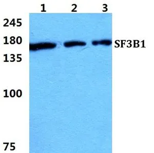
![Mouse tissue extract (50 μg) was separated by 5% SDS-PAGE, and the membrane was blotted with SF3B1 antibody [JB40-32] (GTX01165) diluted at 1:1000. The HRP-conjugated anti-rabbit IgG antibody (GTX213110-01) was used to detect the primary antibody.](https://www.genetex.com/upload/website/prouct_img/normal/GTX01165/GTX01165_HL0222_20200828_WB_M_thymus_w_23053121_505.webp "Mouse tissue extract (50 μg) was separated by 5% SDS-PAGE, and the membrane was blotted with SF3B1 antibody [JB40-32] (GTX01165) diluted at 1:1000. The HRP-conjugated anti-rabbit IgG antibody (GTX213110-01) was used to detect the primary antibody.")
![IHC-P analysis of human colon cancer tissue section using GTX01165 SF3B1 antibody [JB40-32].](https://www.genetex.com/upload/website/prouct_img/normal/GTX01165/GTX01165_20200303_IHC-P_650_w_23053121_431.webp "IHC-P analysis of human colon cancer tissue section using GTX01165 SF3B1 antibody [JB40-32].")
![IHC-P analysis of mouse small intestine tissue section using GTX01165 SF3B1 antibody [JB40-32].](https://www.genetex.com/upload/website/prouct_img/normal/GTX01165/GTX01165_20200303_IHC-P_653_w_23053121_975.webp "IHC-P analysis of mouse small intestine tissue section using GTX01165 SF3B1 antibody [JB40-32].")
IHC-P analysis of human skin tissue section using GTX01165 SF3B1 antibody [JB40-32].
SF3B1 antibody [JB40-32]
GTX01165
ApplicationsWestern Blot, ImmunoHistoChemistry, ImmunoHistoChemistry Paraffin
Product group Antibodies
ReactivityHuman, Mouse, Rat
TargetSF3B1
Overview
- SupplierGeneTex
- Product NameSF3B1 antibody [JB40-32]
- Delivery Days Customer9
- Application Supplier NoteWB: 1:500-1:2000. IHC-P: 1:50-1:200. *Optimal dilutions/concentrations should be determined by the researcher.Not tested in other applications.
- ApplicationsWestern Blot, ImmunoHistoChemistry, ImmunoHistoChemistry Paraffin
- CertificationResearch Use Only
- ClonalityMonoclonal
- Clone IDJB40-32
- Concentration1 mg/ml
- ConjugateUnconjugated
- Gene ID23451
- Target nameSF3B1
- Target descriptionsplicing factor 3b subunit 1
- Target synonymsHsh155, MDS, PRP10, PRPF10, SAP155, SF3b155, splicing factor 3B subunit 1, pre-mRNA processing 10, pre-mRNA splicing factor SF3b, 155 kDa subunit, spliceosome-associated protein 155, splicing factor 3b, subunit 1, 155kDa
- HostRabbit
- IsotypeIgG
- Protein IDO75533
- Protein NameSplicing factor 3B subunit 1
- Scientific DescriptionThis gene encodes subunit 1 of the splicing factor 3b protein complex. Splicing factor 3b, together with splicing factor 3a and a 12S RNA unit, forms the U2 small nuclear ribonucleoproteins complex (U2 snRNP). The splicing factor 3b/3a complex binds pre-mRNA upstream of the introns branch site in a sequence independent manner and may anchor the U2 snRNP to the pre-mRNA. Splicing factor 3b is also a component of the minor U12-type spliceosome. The carboxy-terminal two-thirds of subunit 1 have 22 non-identical, tandem HEAT repeats that form rod-like, helical structures. Alternative splicing results in multiple transcript variants encoding different isoforms. [provided by RefSeq, Jul 2008]
- ReactivityHuman, Mouse, Rat
- Storage Instruction-20°C or -80°C,2°C to 8°C
- UNSPSC41116161
Datasheet
Related products
Product group Antibodies
SF3B1 AntibodyCSB-PA004648
ApplicationsWestern Blot, ELISA
ReactivityHuman, Mouse
TargetSF3B1
- SizePrice
Product group Antibodies
Anti-SF3B1 AntibodyA28151
ApplicationsWestern Blot
ReactivityHuman, Mouse, Rat
- SizePrice

Product group Antibodies
ApplicationsFlow Cytometry, ImmunoFluorescence, Western Blot, ImmunoCytoChemistry, ImmunoHistoChemistry
ReactivityHuman, Mouse, Rat
TargetSF3B1
- SizePrice

Product group Antibodies
Anti-SF3B1 AntibodyHPA050275
ApplicationsWestern Blot, ImmunoCytoChemistry, ImmunoHistoChemistry
ReactivityHuman
TargetSF3B1
- SizePrice
Product group Antibodies

SF3B1 AntibodyLS-C681307
ApplicationsWestern Blot, ELISA, ImmunoHistoChemistry, ImmunoHistoChemistry Paraffin
ReactivityHuman
TargetSF3B1
- SizePrice
Product group Antibodies
SF3B1 Recombinant Antibody, AbBy Fluor-647 ConjugatedBSM-61871R-BF647
ApplicationsImmunoFluorescence, Western Blot
ReactivityHuman, Mouse, Rat
TargetSF3B1
- SizePrice
Product group Antibodies
SF3B1 Polyclonal AntibodyCAC15431
ApplicationsWestern Blot, ELISA, ImmunoHistoChemistry
TargetSF3B1
- SizePrice
Product group Antibodies
SF3B1 antibodyGTX66743
ApplicationsWestern Blot
ReactivityHuman, Mouse, Rat
TargetSF3B1
- SizePrice

Product group Antibodies
SF3B1 antibody, N-termGTX47327
ApplicationsWestern Blot, ImmunoHistoChemistry, ImmunoHistoChemistry Paraffin
ReactivityHuman
TargetSF3B1
- SizePrice