

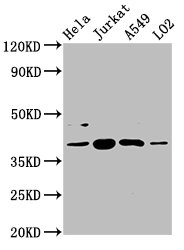
Western Blot Positive WB detected in: Hela whole cell lysate, Jurkat whole cell lysate, A549 whole cell lysate, LO2 whole cell lysate All lanes: SHARPIN antibody at 5.4microg/ml Secondary Goat polyclonal to rabbit IgG at 1/50000 dilution Predicted band size: 40, 34 kDa Observed band size: 40 kDa
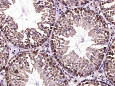
. Section was blocked with 10% normal goat serum 30min at RT. Then primary antibody (1% BSA) was incubated at 4°C overnight. The primary is detected by a biotinylated secondary antibody and visualized using an HRP conjugated SP system.")
. Section was blocked with 10% normal goat serum 30min at RT. Then primary antibody (1% BSA) was incubated at 4°C overnight. The primary is detected by a biotinylated secondary antibody and visualized using an HRP conjugated SP system.")
.")
Western Blot Positive WB detected in: Hela whole cell lysate, Jurkat whole cell lysate, A549 whole cell lysate, LO2 whole cell lysate All lanes: SHARPIN antibody at 5.4microg/ml Secondary Goat polyclonal to rabbit IgG at 1/50000 dilution Predicted band size: 40, 34 kDa Observed band size: 40 kDa
SHARPIN Antibody
CSB-PA867119LA01HU
ApplicationsImmunoFluorescence, Western Blot, ELISA, ImmunoHistoChemistry
Product group Antibodies
ReactivityHuman
TargetSHARPIN
Overview
- SupplierCusabio
- Product NameSHARPIN Antibody
- Delivery Days Customer20
- ApplicationsImmunoFluorescence, Western Blot, ELISA, ImmunoHistoChemistry
- CertificationResearch Use Only
- ClonalityPolyclonal
- ConjugateUnconjugated
- Gene ID81858
- Target nameSHARPIN
- Target descriptionSHANK associated RH domain interactor
- Target synonymsAIFID, SIPL1, sharpin, hSIPL1, shank-associated RH domain-interacting protein, shank-interacting protein-like 1
- HostRabbit
- IsotypeIgG
- Protein IDQ9H0F6
- Protein NameSharpin
- Scientific DescriptionComponent of the LUBAC complex which conjugates linear polyubiquitin chains in a head-to-tail manner to substrates and plays a key role in NF-kappa-B activation and regulation of inflammation. LUBAC conjugates linear polyubiquitin to IKBKG and RIPK1 and is involved in activation of the canonical NF-kappa-B and the JNK signaling pathways. Linear ubiquitination mediated by the LUBAC complex interferes with TNF-induced cell death and thereby prevents inflammation. LUBAC is proposed to be recruited to the TNF-R1 signaling complex (TNF-RSC) following polyubiquitination of TNF-RSC components by BIRC2 and/or BIRC3 and to conjugate linear polyubiquitin to IKBKG and possibly other components contributing to the stability of the complex. Together with FAM105B/otulin, the LUBAC complex regulates the canonical Wnt signaling during angiogenesis.
- ReactivityHuman
- Storage Instruction-20°C or -80°C
- UNSPSC41116161
Related products
Product group Antibodies

Anti-SHARPIN [RAB-S246]Ab01868-1.1
ApplicationsFlow Cytometry
ReactivityHuman
TargetSHARPIN
- SizePrice
Product group Antibodies
Anti-SHARPIN Antibody144-12240
ApplicationsWestern Blot
ReactivityHuman, Rat
TargetSHARPIN
- SizePrice
Product group Antibodies
SHARPIN antibodyGTX130231
ApplicationsImmunoPrecipitation, Western Blot
ReactivityHuman
TargetSHARPIN
- SizePrice
Product group Antibodies
Sharpin Polyclonal AntibodyCAC11354
ApplicationsImmunoFluorescence, Western Blot, ELISA, ImmunoHistoChemistry
TargetSHARPIN
- SizePrice
Product group Antibodies
SHARPIN Polyclonal AntibodyBS-9581R
ApplicationsImmunoFluorescence, Western Blot, ImmunoHistoChemistry, ImmunoHistoChemistry Paraffin
ReactivityCanine, Human, Mouse, Rat
TargetSHARPIN
- SizePrice
Product group Antibodies
Anti-SHARPIN AntibodyA80634
ApplicationsWestern Blot
ReactivityHuman, Rat
- SizePrice

Product group Antibodies
Anti-SHARPIN Antibody Picoband(r)A06687-2-CARRIER-FREE
ApplicationsFlow Cytometry, Western Blot, ELISA, ImmunoHistoChemistry
ReactivityHuman, Mouse, Rat
TargetSHARPIN
- SizePrice
Product group Antibodies

SHARPIN AntibodyLS-C747094
ApplicationsWestern Blot
ReactivityHuman, Rat
TargetSHARPIN
- SizePrice

Product group Antibodies
Anti-SHARPIN AntibodyHPA044453
ApplicationsWestern Blot, ImmunoHistoChemistry
ReactivityHuman
TargetSHARPIN
- SizePrice