

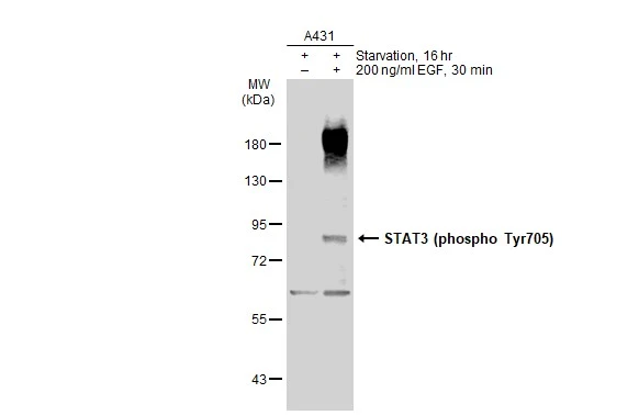
Untreated (–) and treated (+) A431 whole cell extracts (30 μg) were separated by 7.5% SDS-PAGE, and the membrane was blotted with STAT3 (phospho Tyr705) antibody (GTX118000) diluted at 1:500. The HRP-conjugated anti-rabbit IgG antibody (GTX213110-01) was used to detect the primary antibody.
 of paraformaldehyde-fixed A431, using STAT3 (Phospho Y705)(GTX118000) antibody (Green) at 1:500 dilution. Alpha-tubulin filaments were labeled with GTX11304 (Red) at 1:2000.")


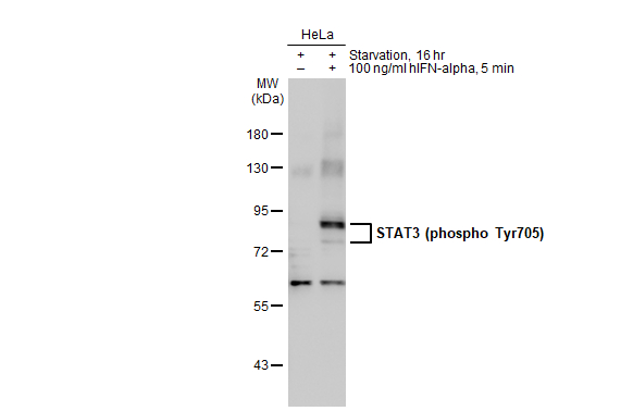
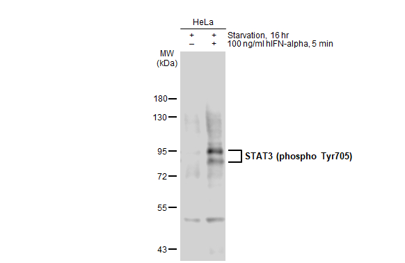
 and treated (+) HeLa whole cell extracts (30 μg) were separated by 7.5% SDS-PAGE, and the membrane was blotted with STAT3 (phospho Tyr705) antibody (GTX118000) diluted at 1:1000. The HRP-conjugated anti-rabbit IgG antibody (GTX213110-01) was used to detect the primary antibody, and the signal was developed with Trident ECL plus-Enhanced.")
Untreated (–) and treated (+) A431 whole cell extracts (30 μg) were separated by 7.5% SDS-PAGE, and the membrane was blotted with STAT3 (phospho Tyr705) antibody (GTX118000) diluted at 1:500. The HRP-conjugated anti-rabbit IgG antibody (GTX213110-01) was used to detect the primary antibody.
STAT3 (phospho Tyr705) antibody
GTX118000
ApplicationsFlow Cytometry, ImmunoFluorescence, Western Blot, ImmunoCytoChemistry, ImmunoHistoChemistry, ImmunoHistoChemistry Paraffin
Product group Antibodies
ReactivityHuman, Mouse, Zebra Fish
TargetSTAT3
Overview
- SupplierGeneTex
- Product NameSTAT3 (phospho Tyr705) antibody
- Delivery Days Customer9
- Application Supplier NoteWB: 1:500-1:3000. ICC/IF: 1:100-1:1000. *Optimal dilutions/concentrations should be determined by the researcher.Not tested in other applications.
- ApplicationsFlow Cytometry, ImmunoFluorescence, Western Blot, ImmunoCytoChemistry, ImmunoHistoChemistry, ImmunoHistoChemistry Paraffin
- CertificationResearch Use Only
- ClonalityPolyclonal
- Concentration1 mg/ml
- ConjugateUnconjugated
- Gene ID6774
- Target nameSTAT3
- Target descriptionsignal transducer and activator of transcription 3
- Target synonymsADMIO, ADMIO1, APRF, HIES, signal transducer and activator of transcription 3, DNA-binding protein APRF, acute-phase response factor
- HostRabbit
- IsotypeIgG
- Protein IDP40763
- Protein NameSignal transducer and activator of transcription 3
- Scientific DescriptionThe protein encoded by this gene is a member of the STAT protein family. In response to cytokines and growth factors, STAT family members are phosphorylated by the receptor associated kinases, and then form homo- or heterodimers that translocate to the cell nucleus where they act as transcription activators. This protein is activated through phosphorylation in response to various cytokines and growth factors including IFNs, EGF, IL5, IL6, HGF, LIF and BMP2. This protein mediates the expression of a variety of genes in response to cell stimuli, and thus plays a key role in many cellular processes such as cell growth and apoptosis. The small GTPase Rac1 has been shown to bind and regulate the activity of this protein. PIAS3 protein is a specific inhibitor of this protein. Three alternatively spliced transcript variants encoding distinct isoforms have been described. [provided by RefSeq]
- ReactivityHuman, Mouse, Zebra Fish
- Storage Instruction-20°C or -80°C,2°C to 8°C
- UNSPSC12352203
References
- Wang BY, Shen HT, Lee YL, et al. Inhibition of Na+/H+ exchanger (NHE) 7 by 5-(N-ethyl-N-isopropyl)-Amiloride displays anti-cancer activity in non-small cell lung cancer by disrupting cancer stem cell activity and downregulating PD-L1 expression. Am J Cancer Res. 2023,13(10):4721-4733.Read this paper
- Zhao M, Zheng Z, Zhang P, et al. IL-30 protects against sepsis-induced myocardial dysfunction by inhibiting pro-inflammatory macrophage polarization and pyroptosis. iScience. 2023,26(9):107544. doi: 10.1016/j.isci.2023.107544Read this paper
- Di Giorgio C, Bellini R, Lupia A, et al. Discovery of BAR502, as potent steroidal antagonist of leukemia inhibitory factor receptor for the treatment of pancreatic adenocarcinoma. Front Oncol. 2023,13:1140730. doi: 10.3389/fonc.2023.1140730Read this paper
- Di Giorgio C, Lupia A, Marchianò S, et al. Repositioning Mifepristone as a Leukaemia Inhibitory Factor Receptor Antagonist for the Treatment of Pancreatic Adenocarcinoma. Cells. 2022,11(21). doi: 10.3390/cells11213482Read this paper
- Di Giorgio C, Marchianò S, Marino E, et al. Next-Generation Sequencing Analysis of Gastric Cancer Identifies the Leukemia Inhibitory Factor Receptor as a Driving Factor in Gastric Cancer Progression and as a Predictor of Poor Prognosis. Front Oncol. 2022,12:939969. doi: 10.3389/fonc.2022.939969Read this paper
- Lin ZH, Hu J, Shi H, et al. Water extract of medicinal ink (WEMI) attenuates lipopolysaccharide-induced NO production of Raw264.7 cells via downregulating JAK2/STAT3-mediated iNOS expression. J Ethnopharmacol. 2022,282:114636. doi: 10.1016/j.jep.2021.114636Read this paper
- Zhang L, Sui C, Zhang Y, et al. Knockdown of hsa_circ_0134111 alleviates the symptom of osteoarthritis via sponging microRNA-224-5p. Cell Cycle. 2021,20(11):1052-1066. doi: 10.1080/15384101.2021.1919838Read this paper
- Wu SY, Yang WY, Cheng CC, et al. Low molecular weight fucoidan inhibits hepatocarcinogenesis and nonalcoholic fatty liver disease in zebrafish via ASGR/STAT3/HNF4A signaling. Clin Transl Med. 2020,10(8):e252. doi: 10.1002/ctm2.252Read this paper
- Castañeda-Zárraga A, Rodríguez-Cid JR, Flores-Mariñelarena RR, et al. Human skin biomarkers relationship to response to treatment with tyrosine kinase inhibitors in advanced EGFR-mutated lung adenocarcinoma. Thorac Cancer. 2020,11(11):3243-3251. doi: 10.1111/1759-7714.13657Read this paper
- Wang YY, Xiao LY, Chen YK, et al. Orabase-Formulated Benzalkonium Chloride Effectively Suppressed Oral Potentially Malignant Disorder In Vitro and In Vivo. ACS Omega. 2020,5(12):7018-7024. doi: 10.1021/acsomega.0c00640Read this paper
Datasheet
Related products
Product group Antibodies

Anti-STAT3 phosphorylated [AbAb1-pSTAT3]AB04100-10.0
ApplicationsImmunoPrecipitation, Western Blot, ELISA
ReactivityHuman, Mouse
TargetSTAT3
- SizePrice
Product group Antibodies
Anti-P-STAT3 Antibody130-10609
ApplicationsELISA
ReactivityHuman
TargetSTAT3
- SizePrice
Product group Antibodies
References
STAT3 (phospho Tyr705) antibodyGTX133464
ApplicationsWestern Blot
ReactivityHuman, Mouse
TargetSTAT3
- SizePrice
Product group Antibodies
STAT3 (phospho Tyr705) antibodyGTX133615
ApplicationsWestern Blot
ReactivityHuman
TargetSTAT3
- SizePrice
![Untreated (–) and treated (+) HeLa whole cell extracts (30 μg) were separated by 7.5% SDS-PAGE, and the membrane was blotted with STAT3 (phospho Tyr705) antibody [GT1204] (GTX00966) diluted at 1:500. The HRP-conjugated anti-rabbit IgG antibody (GTX213110-01) was used to detect the primary antibody.](https://www.genetex.com/upload/website/prouct_img/normal/GTX00966/GTX00966_4000000140_20200313_WB_treatment_hIFN-alpha_w_23053121_556.webp)
Product group Antibodies
ApplicationsWestern Blot
ReactivityHuman, Mouse
TargetSTAT3
- SizePrice
![IHC-P analysis of mouse kidney tissue section using GTX00968 STAT3 (phospho Ser727) antibody [GT1206]. Dilution : 1:100](https://www.genetex.com/upload/website/prouct_img/normal/GTX00968/GTX00968_20200327_IHC-P_34_w_23053121_978.webp)
Product group Antibodies
ApplicationsWestern Blot, ImmunoHistoChemistry, ImmunoHistoChemistry Paraffin
ReactivityHuman, Mouse, Rat
TargetSTAT3
- SizePrice
![Various whole cell extracts (30 μg) were separated by 7.5% SDS-PAGE, and the membrane was blotted with STAT3 antibody [GT1159] (GTX01294) diluted at 1:500. The HRP-conjugated anti-rabbit IgG antibody (GTX213110-01) was used to detect the primary antibody.](https://www.genetex.com/upload/website/prouct_img/normal/GTX01294/GTX01294_40000000045_20200306_WB_w_23053121_409.webp)
Product group Antibodies
STAT3 antibody [GT1159]GTX01294
ApplicationsWestern Blot
ReactivityHuman, Mouse, Rat
TargetSTAT3
- SizePrice
![Various whole cell extracts (30 μg) were separated by 7.5% SDS-PAGE, and the membrane was blotted with STAT3 antibody [C3], C-term (GTX104616) diluted at 1:1000. The HRP-conjugated anti-rabbit IgG antibody (GTX213110-01) was used to detect the primary antibody.](https://www.genetex.com/upload/website/prouct_img/normal/GTX104616/GTX104616_43873_20210226_WB_R_w_23060120_540.webp)
Product group Antibodies
References
STAT3 antibody [C3], C-termGTX104616
ApplicationsImmunoFluorescence, Western Blot, ImmunoCytoChemistry, ImmunoHistoChemistry, ImmunoHistoChemistry Paraffin
ReactivityHuman, Mouse, Plant, Rat
TargetSTAT3
- SizePrice
Product group Antibodies
References
STAT3 antibodyGTX108630
ApplicationsFlow Cytometry, ImmunoPrecipitation, Western Blot
ReactivityHuman, Zebra Fish
TargetSTAT3
- SizePrice