

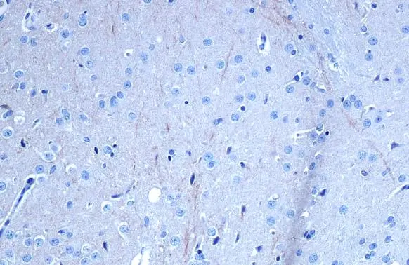
TrkB antibody detects TrkB protein at cell membrane by immunohistochemical analysis. Sample: Paraffin-embedded rat brain. TrkB stained by TrkB antibody (GTX133722) diluted at 1:500. Antigen Retrieval: Citrate buffer, pH 6.0, 15 min
 diluted at 1:500. Antigen Retrieval: Citrate buffer, pH 6.0, 15 min")
![TrkB antibody detects TrkB protein by immunofluorescent analysis. Sample: DIV10 rat E18 primary hippocampal neurons were fixed in 4% paraformaldehyde at RT for 15 min. Green: TrkB protein stained by TrkB antibody (GTX133722) diluted at 1:500. Red: beta Tubulin 3/ Tuj1, stained by beta Tubulin 3/ Tuj1 antibody [GT1338] (GTX631831) diluted at 1:500. Blue: Fluoroshield with DAPI (GTX30920).](https://www.genetex.com/upload/website/prouct_img/normal/GTX133722/GTX133722_42879_20170824_IFA_w_23060523_108.webp "TrkB antibody detects TrkB protein by immunofluorescent analysis. Sample: DIV10 rat E18 primary hippocampal neurons were fixed in 4% paraformaldehyde at RT for 15 min. Green: TrkB protein stained by TrkB antibody (GTX133722) diluted at 1:500. Red: beta Tubulin 3/ Tuj1, stained by beta Tubulin 3/ Tuj1 antibody [GT1338] (GTX631831) diluted at 1:500. Blue: Fluoroshield with DAPI (GTX30920).")
 was separated by 5% SDS-PAGE, and the membranes were blotted with TrkB antibody (GTX133722) diluted at 1:1000 and competitor's antibody (sc-12) diluted at 1:100. The HRP-conjugated anti-rabbit IgG antibody (GTX213110-01) was used to detect the primary antibody.")
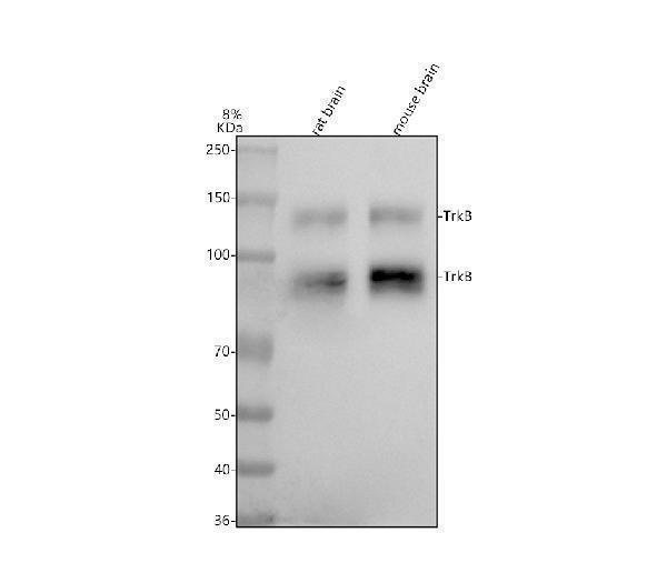
 were separated by 7.5% SDS-PAGE, and the membrane was blotted with TrkB antibody (GTX133722) diluted at 1:1000. The HRP-conjugated anti-rabbit IgG antibody (GTX213110-01) was used to detect the primary antibody.")
TrkB antibody detects TrkB protein at cell membrane by immunohistochemical analysis. Sample: Paraffin-embedded rat brain. TrkB stained by TrkB antibody (GTX133722) diluted at 1:500. Antigen Retrieval: Citrate buffer, pH 6.0, 15 min
TrkB antibody
GTX133722
ApplicationsImmunoFluorescence, Western Blot, ImmunoCytoChemistry, ImmunoHistoChemistry, ImmunoHistoChemistry Paraffin
Product group Antibodies
ReactivityMouse, Rat
TargetNtrk2
Overview
- SupplierGeneTex
- Product NameTrkB antibody
- Delivery Days Customer9
- Application Supplier NoteWB: 1:500-1:3000. ICC/IF: 1:100-1:1000. *Optimal dilutions/concentrations should be determined by the researcher.Not tested in other applications.
- ApplicationsImmunoFluorescence, Western Blot, ImmunoCytoChemistry, ImmunoHistoChemistry, ImmunoHistoChemistry Paraffin
- CertificationResearch Use Only
- ClonalityPolyclonal
- Concentration1.24 mg/ml
- ConjugateUnconjugated
- Gene ID18212
- Target nameNtrk2
- Target descriptionneurotrophic tyrosine kinase, receptor, type 2
- Target synonymsGP145-TrkB/GP95-TrkB, Tkrb, trk-B, trkB, BDNF/NT-3 growth factors receptor, neurotrophic tyrosine receptor kinase type 2, trkB tyrosine kinase
- HostRabbit
- IsotypeIgG
- Protein IDP15209
- Protein NameBDNF/NT-3 growth factors receptor
- Scientific DescriptionReceptor for brain-derived neurotrophic factor (BDNF), neurotrophin-3 and neurotrophin-4/5 but not nerve growth factor (NGF). Involved in the development and/or maintenance of the nervous system. This is a tyrosine-protein kinase receptor. Known substrates for the TRK receptors are SHC1, PI-3 kinase, and PLC-gamma-1.
- ReactivityMouse, Rat
- Storage Instruction-20°C or -80°C,2°C to 8°C
- UNSPSC41116161
Datasheet
Related products
Product group Antibodies

Mouse Ntrk2 AntibodyABX027692
ApplicationsWestern Blot, ELISA, ImmunoHistoChemistry
- SizePrice
Product group Antibodies
References
ApplicationsImmunoPrecipitation, Western Blot, ImmunoHistoChemistry
ReactivityMouse, Rat
TargetNtrk2
- SizePrice
![IHC-P analysis of lung and pancrease tissue sections from LPS exposed mouse using GTX53109 TrkB antibody [6B10].](https://www.genetex.com/upload/website/prouct_img/normal/GTX53109/GTX53109_20191119_IHC-P_w_23060900_434.webp)
Product group Antibodies
TrkB antibody [6B10]GTX53109
ApplicationsWestern Blot, ImmunoHistoChemistry, ImmunoHistoChemistry Paraffin
ReactivityMouse
TargetNtrk2
- SizePrice