

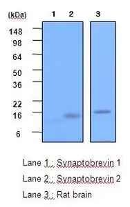
WB analysis of recombinant Synaptobrevin 1, recombinant Synaptobrevin 2 and rat brain lysate (20ug per lane) using Synaptobrevin 2 antibody at a dilution of 1:2,000.
WB analysis of recombinant Synaptobrevin 1, recombinant Synaptobrevin 2 and rat brain lysate (20ug per lane) using Synaptobrevin 2 antibody at a dilution of 1:2,000.
VAMP2 antibody [3E5]
GTX53789
ApplicationsWestern Blot, ELISA
Product group Antibodies
ReactivityHuman, Rat
TargetVAMP2
Overview
- SupplierGeneTex
- Product NameVAMP2 antibody [3E5]
- Delivery Days Customer9
- Application Supplier NoteThe antibody has been tested by ELISA and Western blot analysis to assure specificity and reactivity. Since application varies, however, each investigation should be titrated by the reagent to obtain optimal results. Recommended dilution range for Western blot analysis is 1:1,000 ~ 3,000. Recommended starting dilution is 1:2,000.
- ApplicationsWestern Blot, ELISA
- CertificationResearch Use Only
- ClonalityMonoclonal
- Concentration1 mg/ml
- ConjugateUnconjugated
- Gene ID6844
- Target nameVAMP2
- Target descriptionvesicle associated membrane protein 2
- Target synonymsNEDHAHM, SYB2, VAMP-2, vesicle-associated membrane protein 2, synaptobrevin 2
- HostMouse
- IsotypeIgG1
- Protein IDP63027
- Protein NameVesicle-associated membrane protein 2
- Scientific DescriptionThe protein encoded by this gene is a member of the vesicle-associated membrane protein (VAMP)/synaptobrevin family. Synaptobrevins/VAMPs, syntaxins, and the 25-kD synaptosomal-associated protein SNAP25 are the main components of a protein complex involved in the docking and/or fusion of synaptic vesicles with the presynaptic membrane. This gene is thought to participate in neurotransmitter release at a step between docking and fusion. The protein forms a stable complex with syntaxin, synaptosomal-associated protein, 25 kD, and synaptotagmin. It also forms a distinct complex with synaptophysin. It is a likely candidate gene for familial infantile myasthenia (FIMG) because of its map location and because it encodes a synaptic vesicle protein of the type that has been implicated in the pathogenesis of FIMG. [provided by RefSeq, Jul 2008]
- ReactivityHuman, Rat
- Storage Instruction-20°C or -80°C,2°C to 8°C
- UNSPSC41116161
Datasheet
Related products
Product group Antibodies
Anti-VAMP2 AntibodyA29538
ApplicationsWestern Blot, ImmunoHistoChemistry
ReactivityHuman, Mouse, Rat
- SizePrice
Product group Antibodies
Anti-VAMP2 Antibody144-60082
ApplicationsWestern Blot, ImmunoHistoChemistry
ReactivityHuman, Mouse, Rat
TargetVAMP2
- SizePrice
Product group Antibodies

VAMP2 / VAMP-2 AntibodyLS-C771690
ApplicationsWestern Blot, ELISA, ImmunoHistoChemistry
ReactivityHuman, Mouse, Rat
TargetVAMP2
- SizePrice

Product group Antibodies
VAMP2 Recombinant AntibodyBSM-52925R
ApplicationsFlow Cytometry, ImmunoFluorescence, Western Blot, ImmunoCytoChemistry, ImmunoHistoChemistry, ImmunoHistoChemistry Paraffin
ReactivityHuman, Mouse, Rat
TargetVAMP2
- SizePrice
Product group Antibodies
VAMP2 AntibodyCSB-PA025781ESR2HU
ApplicationsELISA, ImmunoHistoChemistry
ReactivityHuman
TargetVAMP2
- SizePrice

Product group Antibodies
ApplicationsFlow Cytometry, ImmunoFluorescence, ImmunoPrecipitation, Western Blot, ImmunoCytoChemistry
ReactivityHuman, Mouse, Rat
TargetVAMP2
- SizePrice
![VAMP2 antibody detects VAMP2 protein at synaptic vesicles by immunofluorescent analysis. Sample: DIV9 rat E18 primary cortical neurons were fixed in 4% paraformaldehyde at RT for 15 min. Green: VAMP2 protein stained by VAMP2 antibody (GTX121462) diluted at 1:500. Red: beta Tubulin 3/ Tuj1, stained by beta Tubulin 3/ Tuj1 antibody [GT11710] (GTX631836) diluted at 1:500. Blue: Fluoroshield with DAPI (GTX30920).](https://www.genetex.com/upload/website/prouct_img/normal/GTX121462/GTX121462_40513_20170503_IFA_R_w_23060519_797.webp)
Product group Antibodies
VAMP2 antibodyGTX121462
ApplicationsImmunoFluorescence, Western Blot, ImmunoCytoChemistry
ReactivityHuman, Mouse, Rat
TargetVAMP2
- SizePrice
![VAMP2 antibody [GT6311] detects VAMP2 protein by immunofluorescent analysis. Sample: DIV9 rat E18 primary hippocampal neuron cells were fixed in 4% paraformaldehyde at RT for 15 min. Green: VAMP2 stained by VAMP2 antibody [GT6311] (GTX634812) diluted at 1:500. Red: NeuN, stained by NeuN antibody (GTX132974) diluted at 1:1000. Blue: Fluoroshield with DAPI (GTX30920).](https://www.genetex.com/upload/website/prouct_img/normal/GTX634812/GTX634812_43409_20190306_ICC_IF_R_w_23061202_709.webp)
Product group Antibodies
VAMP2 antibody [GT6311]GTX634812
ApplicationsImmunoFluorescence, Western Blot, ImmunoCytoChemistry, ImmunoHistoChemistry, ImmunoHistoChemistry Paraffin
ReactivityHuman, Mouse, Rat
TargetVAMP2
- SizePrice
![Various tissue extracts (50 μg) were separated by 15% SDS-PAGE, and the membrane was blotted with VAMP2 antibody [GT766] (GTX634829) diluted at 1:1000. The HRP-conjugated anti-mouse IgG antibody (GTX213111-01) was used to detect the primary antibody.](https://www.genetex.com/upload/website/prouct_img/normal/GTX634829/GTX634829_43766_20191122_WB_M_R_w_23061202_566.webp)
Product group Antibodies
VAMP2 antibody [GT766]GTX634829
ApplicationsImmunoFluorescence, Western Blot, ImmunoCytoChemistry, ImmunoHistoChemistry, ImmunoHistoChemistry Paraffin
ReactivityHuman, Mouse, Rat
TargetVAMP2
- SizePrice