

Vinculin antibody detects Vinculin protein by western blot analysis. Whole cell extracts (30 μg) was separated by 7.5% SDS-PAGE, and the membrane was blotted with Vinculin antibody (GTX113294) diluted at 1:1000. The HRP-conjugated anti-rabbit IgG antibody (GTX213110-01) was used to detect the primary antibody.
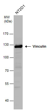
![Mouse tissue extract (50 μg) was separated by 5% SDS-PAGE, and the membrane was blotted with Vinculin antibody [N1N3] (GTX113294) diluted at 1:1000. The HRP-conjugated anti-rabbit IgG antibody (GTX213110-01) was used to detect the primary antibody.](https://www.genetex.com/upload/website/prouct_img/normal/GTX113294/GTX113294_42200_20220218_WB_M_brain_w_23060501_872.webp "Mouse tissue extract (50 μg) was separated by 5% SDS-PAGE, and the membrane was blotted with Vinculin antibody [N1N3] (GTX113294) diluted at 1:1000. The HRP-conjugated anti-rabbit IgG antibody (GTX213110-01) was used to detect the primary antibody.")
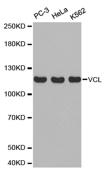
![Various whole cell extracts (30 μg) were separated by 5% SDS-PAGE, and the membrane was blotted with Vinculin antibody [N1N3] (GTX113294) diluted at 1:1000. The HRP-conjugated anti-rabbit IgG antibody (GTX213110-01) was used to detect the primary antibody.](https://www.genetex.com/upload/website/prouct_img/normal/GTX113294/GTX113294_42200_20200306_WB_w_23060501_580.webp "Various whole cell extracts (30 μg) were separated by 5% SDS-PAGE, and the membrane was blotted with Vinculin antibody [N1N3] (GTX113294) diluted at 1:1000. The HRP-conjugated anti-rabbit IgG antibody (GTX213110-01) was used to detect the primary antibody.")
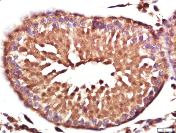
![Vinculin antibody [N1N3] detects Vinculin protein at cytoplasm in mouse testis by immunohistochemical analysis. Sample: Paraffin-embedded mouse testis. Vinculin antibody [N1N3] (GTX113294) diluted at 1:500.
Antigen Retrieval: Citrate buffer, pH 6.0, 15 min](https://www.genetex.com/upload/website/prouct_img/normal/GTX113294/GTX113294_42200_20151013_IHC-P_M_w_23060501_940.webp "Vinculin antibody [N1N3] detects Vinculin protein at cytoplasm in mouse testis by immunohistochemical analysis. Sample: Paraffin-embedded mouse testis. Vinculin antibody [N1N3] (GTX113294) diluted at 1:500.
Antigen Retrieval: Citrate buffer, pH 6.0, 15 min")
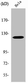
![Various whole cell extracts (30 μg) were separated by 7.5% SDS-PAGE, and the membrane was blotted with Vinculin antibody [N1N3] (GTX113294) diluted at 1:1000. The HRP-conjugated anti-rabbit IgG antibody (GTX213110-01) was used to detect the primary antibody.](https://www.genetex.com/upload/website/prouct_img/normal/GTX113294/GTX113294_42200_20190823_WB_R_w_23060501_134.webp "Various whole cell extracts (30 μg) were separated by 7.5% SDS-PAGE, and the membrane was blotted with Vinculin antibody [N1N3] (GTX113294) diluted at 1:1000. The HRP-conjugated anti-rabbit IgG antibody (GTX213110-01) was used to detect the primary antibody.")
![Vinculin antibody [N1N3] detects Vinculin protein at focal adhesion by immunofluorescent analysis. Sample: HeLa cells were fixed in ice-cold MeOH for 5 min. Green: Vinculin stained by Vinculin antibody [N1N3] (GTX113294) diluted at 1:500. Blue: Fluoroshield with DAPI (GTX30920). Scale bar= 10 μm.](https://www.genetex.com/upload/website/prouct_img/normal/GTX113294/GTX113294_42200_20200219_ICC_IF_w_23060501_219.webp "Vinculin antibody [N1N3] detects Vinculin protein at focal adhesion by immunofluorescent analysis. Sample: HeLa cells were fixed in ice-cold MeOH for 5 min. Green: Vinculin stained by Vinculin antibody [N1N3] (GTX113294) diluted at 1:500. Blue: Fluoroshield with DAPI (GTX30920). Scale bar= 10 μm.")
 antibody at 1:500 dilution.
Antigen Retrieval: Trilogy? (EDTA based, pH 8.0) buffer, 15min")

![Various whole cell extracts (50 μg) were separated by 7.5% SDS-PAGE, and the membrane was blotted with Vinculin antibody [N1N3] (GTX113294) diluted at 1:1000. The HRP-conjugated anti-rabbit IgG antibody (GTX213110-01) was used to detect the primary antibody.](https://www.genetex.com/upload/website/prouct_img/normal/GTX113294/GTX113294_44846_20240503_WB_M_R_24050701_901.webp "Various whole cell extracts (50 μg) were separated by 7.5% SDS-PAGE, and the membrane was blotted with Vinculin antibody [N1N3] (GTX113294) diluted at 1:1000. The HRP-conjugated anti-rabbit IgG antibody (GTX213110-01) was used to detect the primary antibody.")
![Various whole cell extracts (30 μg) were separated by 5% SDS-PAGE, and the membrane was blotted with Vinculin antibody [N1N3] (GTX113294) diluted at 1:10000. The HRP-conjugated anti-rabbit IgG antibody (GTX213110-01) was used to detect the primary antibody.](https://www.genetex.com/upload/website/prouct_img/normal/GTX113294/GTX113294_44846_20240920_WB_24092600_688.webp "Various whole cell extracts (30 μg) were separated by 5% SDS-PAGE, and the membrane was blotted with Vinculin antibody [N1N3] (GTX113294) diluted at 1:10000. The HRP-conjugated anti-rabbit IgG antibody (GTX213110-01) was used to detect the primary antibody.")
Vinculin antibody detects Vinculin protein by western blot analysis. Whole cell extracts (30 μg) was separated by 7.5% SDS-PAGE, and the membrane was blotted with Vinculin antibody (GTX113294) diluted at 1:1000. The HRP-conjugated anti-rabbit IgG antibody (GTX213110-01) was used to detect the primary antibody.
Vinculin antibody [N1N3]
GTX113294
ApplicationsImmunoFluorescence, Western Blot, ImmunoCytoChemistry, ImmunoHistoChemistry, ImmunoHistoChemistry Paraffin
Product group Antibodies
ReactivityHuman, Mouse, Rat
TargetVCL
Overview
- SupplierGeneTex
- Product NameVinculin antibody [N1N3]
- Delivery Days Customer9
- Application Supplier NoteWB: 1:500-1:3000. ICC/IF: 1:100-1:1000. IHC-P: 1:100-1:1000. *Optimal dilutions/concentrations should be determined by the researcher.Not tested in other applications.
- ApplicationsImmunoFluorescence, Western Blot, ImmunoCytoChemistry, ImmunoHistoChemistry, ImmunoHistoChemistry Paraffin
- CertificationResearch Use Only
- ClonalityPolyclonal
- Concentration0.33 mg/ml
- ConjugateUnconjugated
- Gene ID7414
- Target nameVCL
- Target descriptionvinculin
- Target synonymsCMD1W, CMH15, HEL114, MV, MVCL, VINC, vinculin, epididymis luminal protein 114, epididymis secretory sperm binding protein, meta-vinculin, metavinculin
- HostRabbit
- IsotypeIgG
- Protein IDP18206
- Protein NameVinculin
- Scientific DescriptionVinculin is a cytoskeletal protein associated with cell-cell and cell-matrix junctions, where it is thought to function as one of several interacting proteins involved in anchoring F-actin to the membrane. Defects in VCL are the cause of cardiomyopathy dilated type 1W. Dilated cardiomyopathy is a disorder characterized by ventricular dilation and impaired systolic function, resulting in congestive heart failure and arrhythmia. Multiple alternatively spliced transcript variants have been found for this gene, but the biological validity of some variants has not been determined. [provided by RefSeq]
- ReactivityHuman, Mouse, Rat
- Storage Instruction-20°C or -80°C,2°C to 8°C
- UNSPSC41116161
Datasheet
Related products
Product group Antibodies
Anti-VCL AntibodyA39079
ApplicationsWestern Blot, ImmunoHistoChemistry
ReactivityHuman, Mouse, Rat
- SizePrice

Product group Antibodies
Anti-Vinculin/VCL Antibody Picoband(r)A01207-1-CARRIER-FREE
ApplicationsFlow Cytometry, ImmunoFluorescence, Western Blot, ELISA, ImmunoHistoChemistry
ReactivityHuman, Monkey, Mouse, Rat
TargetVCL
- SizePrice
Product group Antibodies
References
Vinculin Polyclonal AntibodyBS-6640R
ApplicationsFlow Cytometry, ImmunoFluorescence, Western Blot, ELISA, ImmunoCytoChemistry, ImmunoHistoChemistry, ImmunoHistoChemistry Frozen, ImmunoHistoChemistry Paraffin
ReactivityBovine, Canine, Chicken, Equine, Human, Mouse, Porcine, Rabbit, Rat
TargetVCL
- SizePrice
Product group Antibodies
VCL AntibodyCSB-PA004452
ApplicationsImmunoFluorescence, Western Blot, ELISA, ImmunoHistoChemistry
ReactivityHuman, Mouse, Rat
TargetVCL
- SizePrice
Product group Antibodies
VCL Polyclonal AntibodyCAC14137
ApplicationsWestern Blot, ELISA
TargetVCL
- SizePrice
![WB analysis of 293, A431, A549, HeLa, HepG2, K562, 3T3, Raji, U937 cell lysate and rat heart, mouse lung, rat lung, mouse spleen, rat spleen, rabbit spleen, mouse brain, rabbit brain and chicken spleen tissue lysate using GTX17308 Vinculin antibody [7E10]. Working concentration : 1 μg/ml](https://www.genetex.com/upload/website/prouct_img/normal/GTX17308/GTX17308_2441_WB_20180221_w_23060620_799.webp)
Product group Antibodies
Vinculin antibody [7E10]GTX17308
ApplicationsWestern Blot, ELISA
ReactivityChicken, Human, Mouse, Rabbit, Rat
TargetVCL
- SizePrice
![WB analysis of U-87 and THP-1 cell lysates using GTX17827 Vinculin antibody [VCL/2572].](https://www.genetex.com/upload/website/prouct_img/normal/GTX17827/GTX17827_20200115_WB_1908_w_23060620_649.webp)
Product group Antibodies
Vinculin antibody [VCL/2572]GTX17827
ApplicationsWestern Blot, ImmunoHistoChemistry, ImmunoHistoChemistry Paraffin, Other Application
ReactivityHuman
TargetVCL
- SizePrice

Product group Antibodies
Anti-VCL AntibodyHPA002131
ApplicationsWestern Blot, ImmunoHistoChemistry
ReactivityHuman, Mouse, Rat
TargetVCL
- SizePrice
![Vinculin antibody [N3C1], Internal detects VCL protein by Western blot analysis. A. 30 μg PC-12 whole cell lysate/extract B. 30 μg Rat2 whole cell lysate/extract 5 % SDS-PAGE Vinculin antibody [N3C1], Internal (GTX109749) dilution: 1:1000](https://www.genetex.com/upload/website/prouct_img/normal/GTX109749/GTX109749_40037_WB_R_w_23060500_998.webp)
Product group Antibodies
Vinculin antibody [N3C1], InternalGTX109749
ApplicationsWestern Blot, ImmunoHistoChemistry, ImmunoHistoChemistry Paraffin
ReactivityHuman, Mouse, Porcine, Rat
TargetVCL
- SizePrice