

VR1 / TRPV1 Antibody (N-Terminus, Sepharose)
LS-C172126
ApplicationsImmunoPrecipitation
Product group Antibodies
TargetTRPV1
Overview
- SupplierLifeSpan BioSciences
- Product NameVR1 / TRPV1 Antibody (N-Terminus, Sepharose)
- Delivery Days Customer23
- ApplicationsImmunoPrecipitation
- Applications SupplierIP
- CertificationResearch Use Only
- ClonalityPolyclonal
- ConjugateSepharose
- Estimated Purity...
- Gene ID7442
- Target nameTRPV1
- Target descriptiontransient receptor potential cation channel subfamily V member 1
- Target synonymscapsaicin receptor; osm-9-like TRP channel 1; OTRPC1; transient receptor potential cation channel subfamily V member 1; transient receptor potential vanilloid 1a; transient receptor potential vanilloid 1b; vanilloid receptor subtype 1; VR1
- HostRabbit
- Storage Instruction2°C to 8°C
- UNSPSC12352203
Related products
Product group Antibodies
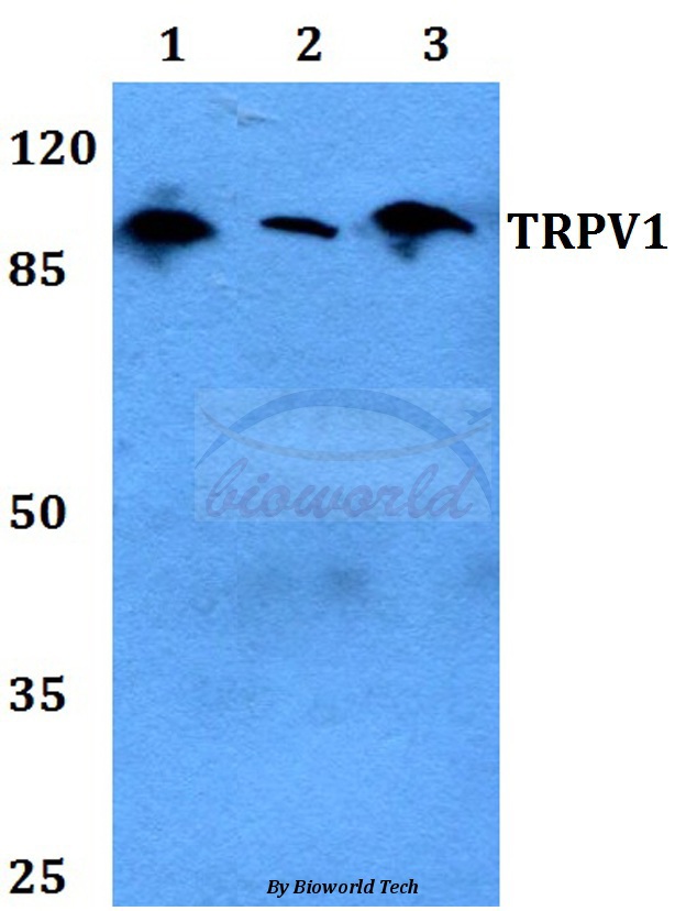
TRPV1 Polyclonal AntibodyCAC13417
ApplicationsImmunoFluorescence, ELISA, ImmunoHistoChemistry
TargetTRPV1
- SizePrice
Product group Antibodies
Anti-TRPV1 Antibody144-66647
ApplicationsImmunoFluorescence, Western Blot
TargetTRPV1
- SizePrice

Product group Antibodies
Anti-TRPV1 Antibody Picoband(r)A00128-4-CARRIER-FREE
ApplicationsFlow Cytometry, ImmunoFluorescence, Western Blot, ELISA, ImmunoCytoChemistry
TargetTRPV1
- SizePrice
Product group Antibodies
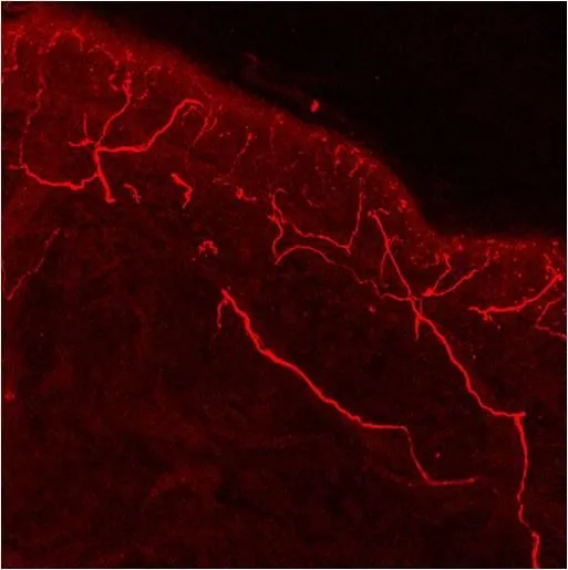
VR1/ TRPV1 antibodyGTX23487
ApplicationsImmunoFluorescence, Western Blot, ImmunoCytoChemistry, ImmunoHistoChemistry, ImmunoHistoChemistry Frozen
TargetTRPV1
- SizePrice

Product group Antibodies
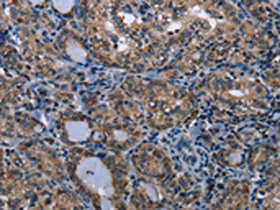
TRPV1 AntibodyCSB-PA037727
ApplicationsELISA, ImmunoHistoChemistry
ReactivityHuman
TargetTRPV1
- SizePrice