

![WT1 (Wilms Tumor 1)(WT1/857 + 6F-H2), Biotin conjugate, 0.1mg/mL [26628-22-8]](https://biotium.com/wp-content/uploads/2016/12/BNUB1138-1-1.jpg "WT1 (Wilms Tumor 1)(WT1/857 + 6F-H2), Biotin conjugate, 0.1mg/mL [26628-22-8]")
![WT1 (Wilms Tumor 1)(WT1/857 + 6F-H2), Biotin conjugate, 0.1mg/mL [26628-22-8]](https://biotium.com/wp-content/uploads/2016/12/BNUB1138-2-1.jpg "WT1 (Wilms Tumor 1)(WT1/857 + 6F-H2), Biotin conjugate, 0.1mg/mL [26628-22-8]")
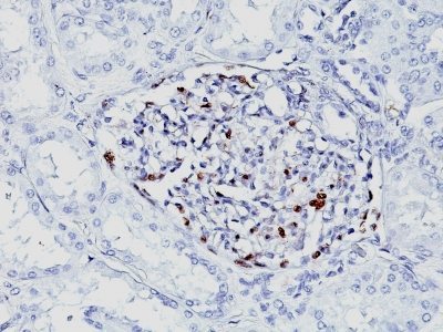
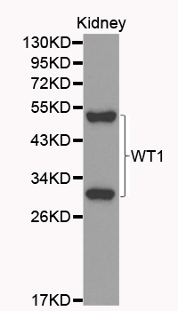
WT1 (Wilms Tumor 1)(WT1/857 + 6F-H2), Biotin conjugate, 0.1mg/mL [26628-22-8]
BNCB1138
ApplicationsImmunoHistoChemistry, ImmunoHistoChemistry Paraffin
Product group Antibodies
ReactivityBovine, Human, Mouse, Rat
TargetWT1
Overview
- SupplierBiotium
- Product NameWT1 (Wilms Tumor 1)(WT1/857 + 6F-H2), Biotin conjugate, 0.1mg/mL [26628-22-8]
- Delivery Days Customer9
- ApplicationsImmunoHistoChemistry, ImmunoHistoChemistry Paraffin
- CAS Number26628-22-8
- CertificationResearch Use Only
- ClonalityMonoclonal
- Clone IDWT1/857 6F-H2
- Concentration0.1 mg/ml
- ConjugateBiotin
- Gene ID7490
- Target nameWT1
- Target descriptionWT1 transcription factor
- Target synonymsAWT1, GUD, NPHS4, WAGR, WIT-2, WT-1, WT33, Wilms tumor protein, Wilms tumor 1
- HostMouse
- IsotypeIgG
- Protein IDP19544
- Protein NameWilms tumor protein
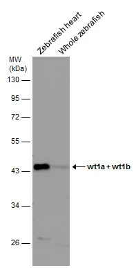
- Scientific DescriptionRecognizes a 47-55 kDa-tumor suppressor protein, identified as Wilms Tumor (WT1) protein. The antibody reacts with all isoforms of the full-length WT1 and also identifies WT1 lacking exon 2-encoded amino acids, frequently found in subsets of sporadic Wilm s tumors.WT1, a sporadic and familial pediatric kidney tumor, is genetically heterogeneous. Wilm s tumor is associated with mutations of WT1, a zinc-finger transcription factor that is essential for the development of the metanephric kidney and the urogenital system. The WT1 gene is normally expressed in fetal kidney and mesothelium, and its expression has been suggested as a marker for Wilm s tumor and mesothelioma. WT1 protein has been identified in proliferative mesothelial cells, malignant mesothelioma, ovarian carcinoma, gonadoblastoma, nephroblastoma, and desmoplastic small round cell tumor. Lung adenocarcinomas rarely stain positive with this antibody. WT1 protein expression in mesothelial cells has become a reliable marker for the diagnosis of mesotheliomas.Primary antibodies are available purified, or with a selection of fluorescent CF® Dyes and other labels. CF® Dyes offer exceptional brightness and photostability. Note: Conjugates of blue fluorescent dyes like CF®405S and CF®405M are not recommended for detecting low abundance targets, because blue dyes have lower fluorescence and can give higher non-specific background than other dye colors.
- SourceAnimal
- ReactivityBovine, Human, Mouse, Rat
- Storage Instruction2°C to 8°C,RT
- UNSPSC41116161
MSDS
Related products
Product group Antibodies
Anti-WT1 AntibodyA35645
ApplicationsImmunoFluorescence, Western Blot, ImmunoHistoChemistry
ReactivityHuman, Mouse, Rat
- SizePrice

Product group Antibodies
Anti-WT1 AntibodyAMAB91839
ApplicationsWestern Blot, ImmunoCytoChemistry, ImmunoHistoChemistry
ReactivityHuman
TargetWT1
- SizePrice

Product group Antibodies
References
ApplicationsFlow Cytometry, ImmunoFluorescence, Western Blot, ImmunoCytoChemistry, ImmunoHistoChemistry
ReactivityHuman, Mouse
TargetWT1
- SizePrice
Product group Antibodies

ApplicationsWestern Blot, ELISA
ReactivityHuman
TargetWT1
- SizePrice
Product group Antibodies
References
WT1 Polyclonal Antibodybs-6983R
ApplicationsFlow Cytometry, ImmunoFluorescence, Western Blot, ELISA, ImmunoCytoChemistry, ImmunoHistoChemistry, ImmunoHistoChemistry Frozen, ImmunoHistoChemistry Paraffin
ReactivityBovine, Canine, Chicken, Human, Mouse, Porcine, Rat, Sheep
TargetWT1
- SizePrice
Product group Antibodies
WT1 AntibodyCSB-PA11609A0RB
ApplicationsELISA, ImmunoHistoChemistry
ReactivityHuman
TargetWT1
- SizePrice
Product group Antibodies

ApplicationsWestern Blot, ELISA
ReactivityHuman
TargetWT1
- SizePrice
Product group Antibodies
ApplicationsImmunoPrecipitation, Western Blot, ImmunoCytoChemistry, ImmunoHistoChemistry
ReactivityMouse, Rat
TargetWT1
- SizePrice
Product group Antibodies
Wilms Tumor 1 antibodyGTX100563
ApplicationsWestern Blot, ImmunoHistoChemistry, ImmunoHistoChemistry Paraffin
ReactivityHuman, Mouse, Rat, Zebra Fish
TargetWT1
- SizePrice