

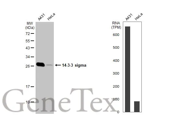
Various whole cell extracts (30 μg) were separated by 12% SDS-PAGE, and the membrane was blotted with 14-3-3 sigma antibody (GTX100289) diluted at 1:3000. The HRP-conjugated anti-rabbit IgG antibody (GTX213110-01) was used to detect the primary antibody. Corresponding RNA expression data for the same cell lines are based on Human Protein Atlas program.
 dilution: 1:3000 The HRP-conjugated anti-rabbit IgG antibody (GTX213110-01) was used to detect the primary antibody.")
 antibody at 1:100 dilution.
Antigen Retrieval: Trilogy? (EDTA based, pH 8.0) buffer, 15min")
 was separated by 12% SDS-PAGE, and the membrane was blotted with 14-3-3 sigma antibody (GTX100289) diluted at 1:3000. The HRP-conjugated anti-rabbit IgG antibody (GTX213110-01) was used to detect the primary antibody.")
 dilution: 1:3000 The HRP-conjugated anti-rabbit IgG antibody (GTX213110-01) was used to detect the primary antibody.")
 dilution: 1:3000 The HRP-conjugated anti-rabbit IgG antibody (GTX213110-01) was used to detect the primary antibody.")
![14-3-3 sigma antibody [C1C3] detects 14-3-3 sigma protein at cytoplasm by immunofluorescent analysis. Sample: A431 cells were fixed in ice-cold Methanol for 5 min. Green: 14-3-3 sigma protein stained by 14-3-3 sigma antibody [C1C3] (GTX100289) diluted at 1:500. Blue: Hoechst 33343 staining.](https://www.genetex.com/upload/website/prouct_img/normal/GTX100289/GTX100289_39456_IFA_w_23060100_137.webp "14-3-3 sigma antibody [C1C3] detects 14-3-3 sigma protein at cytoplasm by immunofluorescent analysis. Sample: A431 cells were fixed in ice-cold Methanol for 5 min. Green: 14-3-3 sigma protein stained by 14-3-3 sigma antibody [C1C3] (GTX100289) diluted at 1:500. Blue: Hoechst 33343 staining.")
 dilution: 1:500.
Antigen Retrieval: Trilogy? (EDTA based, pH 8.0) buffer, 15min")
Various whole cell extracts (30 μg) were separated by 12% SDS-PAGE, and the membrane was blotted with 14-3-3 sigma antibody (GTX100289) diluted at 1:3000. The HRP-conjugated anti-rabbit IgG antibody (GTX213110-01) was used to detect the primary antibody. Corresponding RNA expression data for the same cell lines are based on Human Protein Atlas program.
14-3-3 sigma antibody
GTX100289
ApplicationsImmunoFluorescence, Western Blot, ImmunoCytoChemistry, ImmunoHistoChemistry, ImmunoHistoChemistry Paraffin
Product group Antibodies
ReactivityHuman, Mouse, Rat
TargetSFN
Overview
- SupplierGeneTex
- Product Name14-3-3 sigma antibody
- Delivery Days Customer9
- Application Supplier NoteWB: 1:500-1:3000. ICC/IF: 1:100-1:1000. IHC-P: 1:100-1:1000. *Optimal dilutions/concentrations should be determined by the researcher.Not tested in other applications.
- ApplicationsImmunoFluorescence, Western Blot, ImmunoCytoChemistry, ImmunoHistoChemistry, ImmunoHistoChemistry Paraffin
- CertificationResearch Use Only
- ClonalityPolyclonal
- Concentration1 mg/ml
- ConjugateUnconjugated
- Gene ID2810
- Target nameSFN
- Target descriptionstratifin
- Target synonymsYWHAS, 14-3-3 protein sigma, epithelial cell marker protein 1
- HostRabbit
- IsotypeIgG
- Protein IDP31947
- Protein Name14-3-3 protein sigma
- Scientific DescriptionAdapter protein implicated in the regulation of a large spectrum of both general and specialized signaling pathway. Binds to a large number of partners, usually by recognition of a phosphoserine or phosphothreonine motif. Binding generally results in the modulation of the activity of the binding partner. When bound to KRT17, regulates protein synthesis and epithelial cell growth by stimulating Akt/mTOR pathway. p53-regulated inhibitor of G2/M progression.
- ReactivityHuman, Mouse, Rat
- Storage Instruction-20°C or -80°C,2°C to 8°C
- UNSPSC41116161
Datasheet
Related products
Product group Antibodies
ApplicationsWestern Blot, ELISA
ReactivityHuman, Mouse
- SizePrice

Product group Antibodies
Anti-14-3-3 sigma/SFN Antibody Picoband(r)A01127-CARRIER-FREE
ApplicationsFlow Cytometry, ImmunoFluorescence, Western Blot, ELISA, ImmunoCytoChemistry, ImmunoHistoChemistry
ReactivityHuman, Mouse, Rat
TargetSFN
- SizePrice
Product group Antibodies
Anti-SFN Antibody144-01026
ApplicationsImmunoFluorescence, Western Blot, ImmunoHistoChemistry
ReactivityHuman, Mouse, Rat
TargetSFN
- SizePrice
Product group Antibodies

SFN / Stratifin / 14-3-3 Sigma AntibodyLS-C830366
ApplicationsWestern Blot, ELISA, ImmunoHistoChemistry
ReactivityHuman, Mouse
TargetSFN
- SizePrice
Product group Antibodies
14-3-3 sigma Recombinant AntibodyBSM-60449M
ApplicationsImmunoFluorescence, Western Blot, ImmunoHistoChemistry, ImmunoHistoChemistry Frozen, ImmunoHistoChemistry Paraffin
ReactivityHuman
TargetSFN
- SizePrice
Product group Antibodies
SFN AntibodyCSB-PA008892
ApplicationsWestern Blot, ELISA
ReactivityHuman, Mouse
TargetSFN
- SizePrice
Product group Antibodies

ApplicationsWestern Blot, ELISA, ImmunoHistoChemistry
ReactivityCanine, Human, Mouse, Porcine, Rat
TargetSFN
- SizePrice
Product group Antibodies
SFN Polyclonal AntibodyCAC15119
ApplicationsImmunoFluorescence, Western Blot, ELISA, ImmunoHistoChemistry
ReactivityMouse, Rat
TargetSFN
- SizePrice
![IHC-P analysis of human skin tissue using GTX14116 14-3-3 sigma antibody [1.N.6] (Azide free).](https://www.genetex.com/upload/website/prouct_img/normal/GTX14116/GTX14116_20191203_IHC-P_18_w_23060620_390.webp)
Product group Antibodies
ApplicationsImmunoFluorescence, ImmunoPrecipitation, Western Blot, ImmunoCytoChemistry, ImmunoHistoChemistry, ImmunoHistoChemistry Paraffin
ReactivityHuman
TargetSFN
- SizePrice
![WB analysis of A431 cell lysate using GTX14122 14-3-3 sigma antibody [1.T.28]. Dilution : 0.5μg/ml](https://www.genetex.com/upload/website/prouct_img/normal/GTX14122/GTX14122_20191203_WB_36_w_23060620_216.webp)
Product group Antibodies
14-3-3 sigma antibody [1.T.28]GTX14122
ApplicationsImmunoPrecipitation, Western Blot
ReactivityHuman, Mouse, Rat
TargetSFN
- SizePrice