

4-Aminobutyrate Aminotransferase (ABAT) Polyclonal Antibody
CAU23031
ApplicationsImmunoPrecipitation, Western Blot, ImmunoCytoChemistry, ImmunoHistoChemistry
Product group Antibodies
ReactivityMouse, Rat
TargetABAT
Overview
- SupplierBiomatik
- Product Name4-Aminobutyrate Aminotransferase (ABAT) Polyclonal Antibody
- Delivery Days Customer12
- ApplicationsImmunoPrecipitation, Western Blot, ImmunoCytoChemistry, ImmunoHistoChemistry
- Applications SupplierWB; IHC; ICC; IP.
- CertificationResearch Use Only
- ClonalityPolyclonal
- Concentration0.5 mg/ml
- ConjugateUnconjugated
- Gene ID18
- Target nameABAT
- Target description4-aminobutyrate aminotransferase
- Target synonymsGABA-AT, GABAT, NPD009, 4-aminobutyrate aminotransferase, mitochondrial, (S)-3-amino-2-methylpropionate transaminase, 4-aminobutyrate transaminase, GABA aminotransferase, GABA transaminase, GABA transferase, gamma-amino-N-butyrate transaminase, gamma-aminobutyrate aminotransferase
- HostRabbit
- Protein IDP80404
- Protein Name4-aminobutyrate aminotransferase, mitochondrial
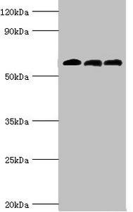
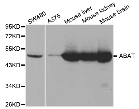
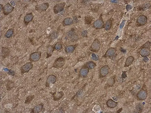
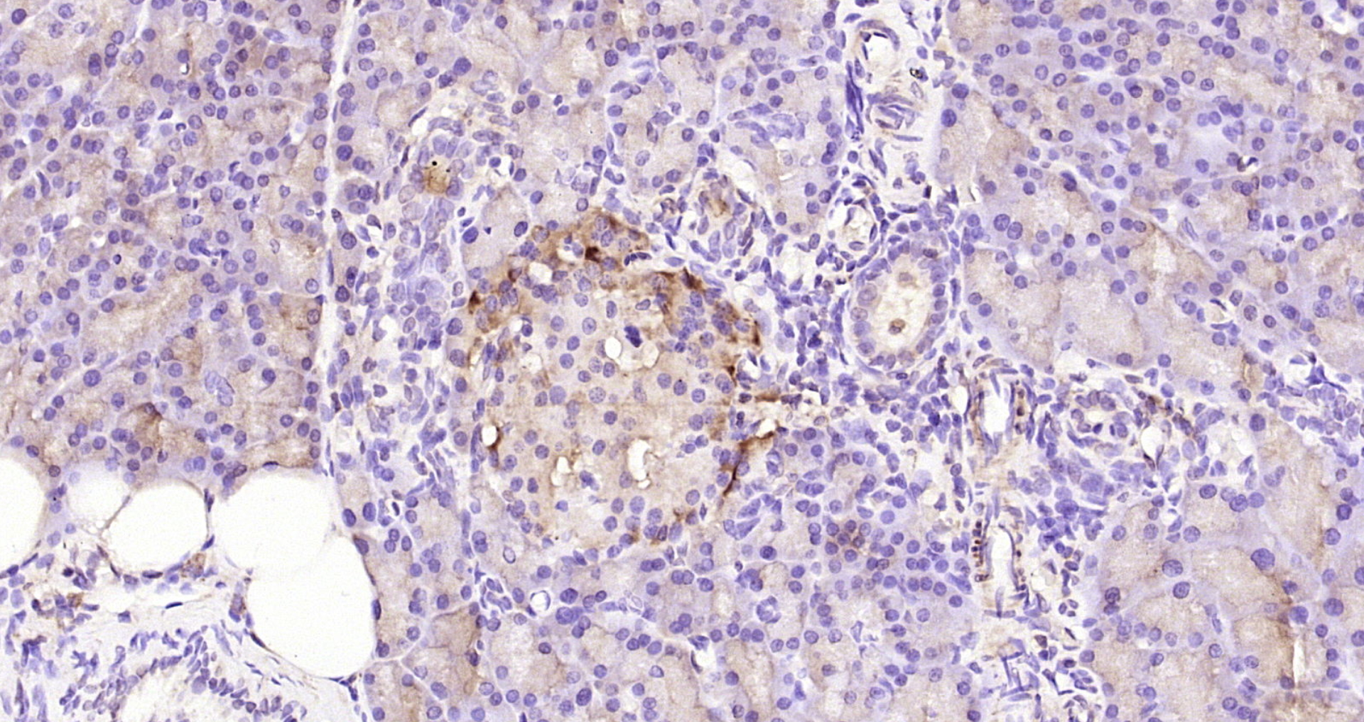
- Scientific DescriptionThe 4-Aminobutyrate Aminotransferase (ABAT) Polyclonal Antibody (Species: Human) has been validated for the following applications: WB, IHC, ICC, IP.
- ReactivityMouse, Rat
- Reactivity SupplierHuman
- Storage Instruction-20°C,2°C to 8°C
- UNSPSC12352203
Related products
Product group Antibodies
ABAT AntibodyCSB-PA001032ESR1HU
ApplicationsWestern Blot, ELISA, ImmunoHistoChemistry
ReactivityHuman, Mouse, Rat
TargetABAT
- SizePrice
Product group Antibodies
Anti-ABAT AntibodyA30938
ApplicationsImmunoFluorescence, Western Blot, ImmunoHistoChemistry
ReactivityHuman, Mouse, Rat
- SizePrice

Product group Antibodies
Anti-ABAT AntibodyHPA041690
ApplicationsWestern Blot, ImmunoHistoChemistry
ReactivityHuman
TargetABAT
- SizePrice
Product group Antibodies

ABAT AntibodyLS-C482413
ApplicationsImmunoFluorescence, Western Blot, ImmunoCytoChemistry, ImmunoHistoChemistry, ImmunoHistoChemistry Paraffin
ReactivityHuman, Mouse, Rat
TargetABAT
- SizePrice

Product group Antibodies
Anti-ABAT Antibody Picoband(r)PB10019-CARRIER-FREE
ApplicationsFlow Cytometry, ImmunoFluorescence, Western Blot, ImmunoCytoChemistry
ReactivityBovine, Human, Mouse, Rat
TargetABAT
- SizePrice
Product group Antibodies
ABAT antibodyGTX100479
ApplicationsImmunoFluorescence, Western Blot, ImmunoCytoChemistry, ImmunoHistoChemistry, ImmunoHistoChemistry Frozen, ImmunoHistoChemistry Paraffin
ReactivityHuman, Mouse, Rat
TargetABAT
- SizePrice
Product group Antibodies
Anti-ABAT Antibody144-05299
ApplicationsImmunoFluorescence, Western Blot, ImmunoHistoChemistry
ReactivityHuman, Mouse, Rat
TargetABAT
- SizePrice
Product group Antibodies
ABAT Polyclonal AntibodyBS-4234R
ApplicationsImmunoFluorescence, Western Blot, ELISA, ImmunoCytoChemistry, ImmunoHistoChemistry, ImmunoHistoChemistry Frozen, ImmunoHistoChemistry Paraffin
ReactivityMouse, Rat
TargetABAT
- SizePrice