

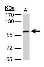
Sample (30μg whole cell lysate) A:A431(GTX27909) 7.5% SDS PAGE GTX100031 diluted at 1:1000
![ABR antibody [C3], C-term detects ABR protein at cell membrane and cytoplasm by immunofluorescent analysis. Sample: A431 cells were fixed in 4% paraformaldehyde at RT for 15 min. Green: ABR protein stained by ABR antibody [C3], C-term (GTX100031) diluted at 1:500. Blue: Hoechst 33342 staining. Scale bar = 10 μm.](https://www.genetex.com/upload/website/prouct_img/normal/GTX100031/GTX100031_39414_20160303_IFA_w_23053123_783.webp "ABR antibody [C3], C-term detects ABR protein at cell membrane and cytoplasm by immunofluorescent analysis. Sample: A431 cells were fixed in 4% paraformaldehyde at RT for 15 min. Green: ABR protein stained by ABR antibody [C3], C-term (GTX100031) diluted at 1:500. Blue: Hoechst 33342 staining. Scale bar = 10 μm.")
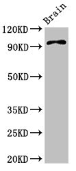
![ABR antibody [C3], C-term detects ABR protein by Western blot analysis. A. 50 μg mouse brain lysate/extract 7.5 % SDS-PAGE ABR antibody [C3], C-term (GTX100031) dilution: 1:1000](https://www.genetex.com/upload/website/prouct_img/normal/GTX100031/GTX100031_39414_WB_M_brain_w_23053123_902.webp "ABR antibody [C3], C-term detects ABR protein by Western blot analysis. A. 50 μg mouse brain lysate/extract 7.5 % SDS-PAGE ABR antibody [C3], C-term (GTX100031) dilution: 1:1000")
Sample (30μg whole cell lysate) A:A431(GTX27909) 7.5% SDS PAGE GTX100031 diluted at 1:1000
ABR antibody [C3], C-term
GTX100031
ApplicationsImmunoFluorescence, Western Blot, ImmunoCytoChemistry
Product group Antibodies
ReactivityHuman, Mouse
TargetABR
Overview
- SupplierGeneTex
- Product NameABR antibody [C3], C-term
- Delivery Days Customer9
- Application Supplier NoteWB: 1:500-1:3000. ICC/IF: 1:100-1:1000. *Optimal dilutions/concentrations should be determined by the researcher.Not tested in other applications.
- ApplicationsImmunoFluorescence, Western Blot, ImmunoCytoChemistry
- CertificationResearch Use Only
- ClonalityPolyclonal
- Concentration1 mg/ml
- ConjugateUnconjugated
- Gene ID29
- Target nameABR
- Target descriptionABR activator of RhoGEF and GTPase
- Target synonymsMDB, active breakpoint cluster region-related protein, ABR, RhoGEF and GTPase activating protein, active BCR-related
- HostRabbit
- IsotypeIgG
- Protein IDQ12979
- Protein NameActive breakpoint cluster region-related protein
- Scientific DescriptionThis gene encodes a protein that is similar to the protein encoded by the breakpoint cluster region gene located on chromosome 22. The protein encoded by this gene contains a GTPase-activating protein domain, a domain found in members of the Rho family of GTP-binding proteins. Functional studies in mice determined that this protein plays a role in vestibular morphogenesis, suggesting that Rho-related GTPases help coordinate motor skills and balance. Alternatively spliced transcript variants that encode different isoforms have been reported for this gene. [provided by RefSeq]
- ReactivityHuman, Mouse
- Storage Instruction-20°C or -80°C,2°C to 8°C
- UNSPSC41116161
Datasheet
Related products
Product group Antibodies
ABR AntibodyCSB-PA622521LA01HU
ApplicationsWestern Blot, ELISA, ImmunoHistoChemistry
ReactivityHuman, Mouse
TargetABR
- SizePrice

Product group Antibodies
Anti-ABR Antibody Picoband(r)A01719-CARRIER-FREE
ApplicationsFlow Cytometry, ImmunoFluorescence, Western Blot, ELISA, ImmunoCytoChemistry, ImmunoHistoChemistry
ReactivityHuman, Mouse, Rat
TargetABR
- SizePrice
Product group Antibodies
Anti-ABR AntibodyA87832
ApplicationsWestern Blot
ReactivityMouse, Rat
- SizePrice
Product group Antibodies

ABR AntibodyLS-C831125
ApplicationsELISA, ImmunoHistoChemistry
ReactivityHuman, Mouse
TargetABR
- SizePrice

Product group Antibodies
Anti-ABR AntibodyHPA053618
ApplicationsImmunoCytoChemistry
ReactivityHuman
TargetABR
- SizePrice
Product group Antibodies
ABR Polyclonal AntibodyCAC15012
ApplicationsWestern Blot, ELISA, ImmunoHistoChemistry
ReactivityMouse
TargetABR
- SizePrice
Product group Antibodies
Anti-ABR Antibody144-63849
ApplicationsWestern Blot
ReactivityHuman, Mouse, Rat
TargetABR
- SizePrice