

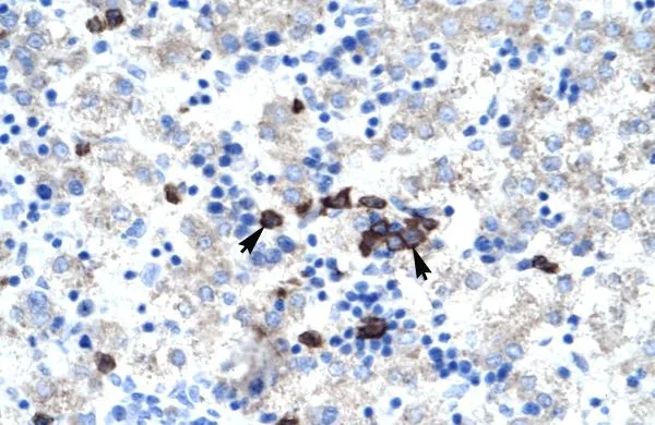
IHC-P analysis of human kidney tissue using GTX16577 ABT1 antibody at 4.0-8.0μg/ml.

IHC-P analysis of human kidney tissue using GTX16577 ABT1 antibody at 4.0-8.0μg/ml.
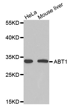
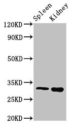
ABT1 antibody, Internal
GTX16577
ApplicationsWestern Blot, ImmunoHistoChemistry, ImmunoHistoChemistry Paraffin
Product group Antibodies
ReactivityHuman
TargetABT1
Overview
- SupplierGeneTex
- Product NameABT1 antibody, Internal
- Delivery Days Customer9
- Application Supplier NoteWB: 0.2-2.5 ug/ml. IHC-P: 2-10 ug/ml. *Optimal dilutions/concentrations should be determined by the researcher.Not tested in other applications.
- ApplicationsWestern Blot, ImmunoHistoChemistry, ImmunoHistoChemistry Paraffin
- CertificationResearch Use Only
- ClonalityPolyclonal
- Concentration0.5-1 mg/ml
- ConjugateUnconjugated
- Gene ID29777
- Target nameABT1
- Target descriptionactivator of basal transcription 1
- Target synonymsEsf2, hABT1, activator of basal transcription 1, TATA-binding protein-binding protein, basal transcriptional activator
- HostRabbit
- IsotypeIgG
- Protein IDQ9ULW3
- Protein NameActivator of basal transcription 1
- Scientific DescriptionBasal transcription of genes by RNA polymerase II requires the interaction of TATA-binding protein (TBP) with the core region of class II promoters. Studies in mouse suggest that the protein encoded by this gene likely activates basal transcription from class II promoters by interaction with TBP and the class II promoter DNA. [provided by RefSeq, Jul 2008]
- ReactivityHuman
- Storage Instruction-20°C or -80°C,2°C to 8°C
- UNSPSC41116161
Datasheet
Related products
Product group Antibodies
Anti-ABT1 AntibodyA38585
ApplicationsWestern Blot, ImmunoHistoChemistry
ReactivityHuman
- SizePrice

Product group Antibodies
ApplicationsWestern Blot
ReactivityHuman
TargetABT1
- SizePrice
Product group Antibodies
ABT1 AntibodyCSB-PA001114LA01HU
ApplicationsImmunoFluorescence, Western Blot, ELISA, ImmunoHistoChemistry
ReactivityHuman, Mouse
TargetABT1
- SizePrice
Product group Antibodies
Abt1 Polyclonal AntibodyCAC08205
ApplicationsImmunoFluorescence, Western Blot, ELISA, ImmunoHistoChemistry
ReactivityMouse
TargetABT1
- SizePrice
Product group Antibodies

HABT1 / ABT1 AntibodyLS-C334372
ApplicationsWestern Blot
ReactivityHuman, Mouse, Rat
TargetABT1
- SizePrice

Product group Antibodies
Anti-ABT1 AntibodyHPA077039
ApplicationsChIP Chromatin ImmunoPrecipitation, ImmunoCytoChemistry
ReactivityHuman
TargetABT1
- SizePrice
Product group Antibodies
Anti-ABT1Y158155
ApplicationsWestern Blot, ELISA, ImmunoHistoChemistry
ReactivityHuman, Mouse, Rat
- SizePrice