

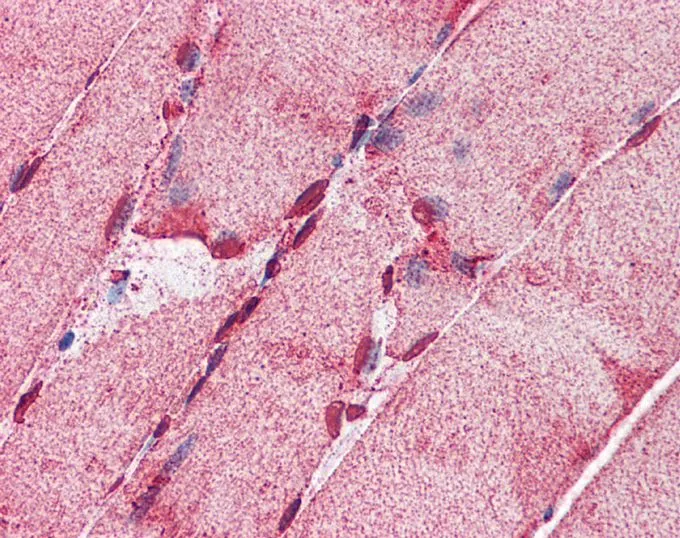
IHC-P analysis of human skeletal muscle tissue using GTX16578 ACSL5 antibody at 5μg/ml.

IHC-P analysis of human skeletal muscle tissue using GTX16578 ACSL5 antibody at 5μg/ml.
ACSL5 antibody, C-term
GTX16578
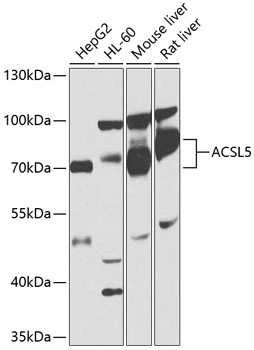
ApplicationsWestern Blot, ImmunoHistoChemistry, ImmunoHistoChemistry Paraffin
Product group Antibodies
ReactivityHuman
TargetACSL5
Overview
- SupplierGeneTex
- Product NameACSL5 antibody, C-term
- Delivery Days Customer9
- Application Supplier NoteWB: 0.2-2.5 ug/ml. IHC-P: 2-10 ug/ml. *Optimal dilutions/concentrations should be determined by the researcher.Not tested in other applications.
- ApplicationsWestern Blot, ImmunoHistoChemistry, ImmunoHistoChemistry Paraffin
- CertificationResearch Use Only
- ClonalityPolyclonal
- Concentration0.5-1 mg/ml
- ConjugateUnconjugated
- Gene ID51703
- Target nameACSL5
- Target descriptionacyl-CoA synthetase long chain family member 5
- Target synonymsACS2, ACS5, DIAR13, FACL5, long-chain-fatty-acid--CoA ligase 5, FACL5 for fatty acid coenzyme A ligase 5, LACS 5, arachidonate--CoA ligase, fatty acid coenzyme A ligase 5, fatty-acid-Coenzyme A ligase, long-chain 5, long-chain acyl-CoA synthetase 5, long-chain fatty acid coenzyme A ligase 5
- HostRabbit
- IsotypeIgG
- Protein IDQ9ULC5
- Protein NameLong-chain-fatty-acid--CoA ligase 5
- Scientific DescriptionThe protein encoded by this gene is an isozyme of the long-chain fatty-acid-coenzyme A ligase family. Although differing in substrate specificity, subcellular localization, and tissue distribution, all isozymes of this family convert free long-chain fatty acids into fatty acyl-CoA esters, and thereby play a key role in lipid biosynthesis and fatty acid degradation. This isozyme is highly expressed in uterus and spleen, and in trace amounts in normal brain, but has markedly increased levels in malignant gliomas. This gene functions in mediating fatty acid-induced glioma cell growth. Three transcript variants encoding two different isoforms have been found for this gene. [provided by RefSeq, Jul 2008]
- ReactivityHuman
- Storage Instruction-20°C or -80°C,2°C to 8°C
- UNSPSC41116161
Datasheet
Related products

Product group Antibodies
Anti-ACSL5 Antibody Picoband(r)A05087-2-CARRIER-FREE
ApplicationsFlow Cytometry, Western Blot
ReactivityHuman, Mouse, Rat
TargetACSL5
- SizePrice
Product group Antibodies
Anti-ACSL5 AntibodyA32127
ApplicationsWestern Blot, ImmunoHistoChemistry
ReactivityHuman
- SizePrice
Product group Antibodies

ACS5 / ACSL5 AntibodyLS-C749143
ApplicationsWestern Blot
ReactivityHuman, Mouse, Rat
TargetACSL5
- SizePrice
Product group Antibodies

Goat anti-ACSL5EB07250
ApplicationsWestern Blot, ELISA, ImmunoHistoChemistry
ReactivityHuman
TargetACSL5
- SizePrice

Product group Antibodies
Anti-ACSL5 AntibodyHPA007162
ApplicationsWestern Blot, ImmunoCytoChemistry, ImmunoHistoChemistry
ReactivityHuman
TargetACSL5
- SizePrice

Product group Antibodies
ACSL5 AntibodyCSB-PA891734HA01HU
ApplicationsWestern Blot, ELISA, ImmunoHistoChemistry
ReactivityHuman, Rat
TargetACSL5
- SizePrice
Product group Antibodies
ACSL5 antibodyGTX30019
ApplicationsWestern Blot, ImmunoHistoChemistry, ImmunoHistoChemistry Paraffin
ReactivityHuman, Mouse, Rat
TargetACSL5
- SizePrice
Product group Antibodies
Acsl5 Polyclonal AntibodyCAC09942
ApplicationsWestern Blot, ELISA, ImmunoHistoChemistry
ReactivityRat
TargetACSL5
- SizePrice
Product group Antibodies
ACSL5 antibody, C-termGTX89348
ApplicationsWestern Blot, ImmunoHistoChemistry, ImmunoHistoChemistry Paraffin
ReactivityHuman
TargetACSL5
- SizePrice