

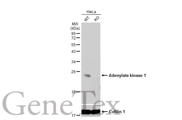
Wild-type (WT) and Adenylate kinase 1 knockout (KO) HeLa cell extracts (30 μg) were separated by 12% SDS-PAGE, and the membrane was blotted with Adenylate kinase 1 antibody [N1C3] (GTX106079) diluted at 1:500. The HRP-conjugated anti-rabbit IgG antibody (GTX213110-01) was used to detect the primary antibody.
 A: Mouse brown adipose tissue 12% SDS PAGE GTX106079 diluted at 1:1000")
![Adenylate kinase 1 antibody [N1C3] detects Adenylate kinase 1 protein at cytosol on Ca922 xenograft by immunohistochemical analysis. Sample: Paraffin-embedded Ca922 xenograft. Adenylate kinase 1 antibody [N1C3] (GTX106079) dilution: 1:500.
Antigen Retrieval: Trilogy? (EDTA based, pH 8.0) buffer, 15min](https://www.genetex.com/upload/website/prouct_img/normal/GTX106079/GTX106079_39890_IHC_w_23060120_329.webp "Adenylate kinase 1 antibody [N1C3] detects Adenylate kinase 1 protein at cytosol on Ca922 xenograft by immunohistochemical analysis. Sample: Paraffin-embedded Ca922 xenograft. Adenylate kinase 1 antibody [N1C3] (GTX106079) dilution: 1:500.
Antigen Retrieval: Trilogy? (EDTA based, pH 8.0) buffer, 15min")
Wild-type (WT) and Adenylate kinase 1 knockout (KO) HeLa cell extracts (30 μg) were separated by 12% SDS-PAGE, and the membrane was blotted with Adenylate kinase 1 antibody [N1C3] (GTX106079) diluted at 1:500. The HRP-conjugated anti-rabbit IgG antibody (GTX213110-01) was used to detect the primary antibody.
Adenylate kinase 1 antibody [N1C3]
GTX106079
ApplicationsWestern Blot, ImmunoHistoChemistry, ImmunoHistoChemistry Paraffin
Product group Antibodies
ReactivityHuman, Mouse
TargetAK1
Overview
- SupplierGeneTex
- Product NameAdenylate kinase 1 antibody [N1C3]
- Delivery Days Customer9
- Application Supplier NoteWB: 1:500-1:3000. IHC-P: 1:100-1:1000. *Optimal dilutions/concentrations should be determined by the researcher.Not tested in other applications.
- ApplicationsWestern Blot, ImmunoHistoChemistry, ImmunoHistoChemistry Paraffin
- CertificationResearch Use Only
- ClonalityPolyclonal
- Concentration0.4 mg/ml
- ConjugateUnconjugated
- Gene ID203
- Target nameAK1
- Target descriptionadenylate kinase 1
- Target synonymsADK, Adk1, CNSHA3, HTL-S-58j, adenylate kinase isoenzyme 1, ATP-AMP transphosphorylase 1, ATP:AMP phosphotransferase, adenylate monophosphate kinase, epididymis secretory sperm binding protein, myokinase, testis secretory sperm binding protein Li 58j
- HostRabbit
- IsotypeIgG
- Protein IDP00568
- Protein NameAdenylate kinase isoenzyme 1
- Scientific DescriptionAdenylate kinase is an enzyme involved in regulating the adenine nucleotide composition within a cell by catalyzing the reversible transfer of phosphate group among adinine nucleotides. Three isozymes of adenylate kinase have been identified in vertebrates, adenylate isozyme 1 (AK1), 2 (AK2) and 3 (AK3). AK1 is found in the cytosol of skeletal muscle, brain and erythrocytes, whereas AK2 and AK3 are found in the mitochondria of other tissues including liver and heart. AK1 was identified because of its association with a rare genetic disorder causing nonspherocytic hemolytic anemia where a mutation in the AK1 gene was found to reduce the catalytic activity of the enzyme. [provided by RefSeq]
- ReactivityHuman, Mouse
- Storage Instruction-20°C or -80°C,2°C to 8°C
- UNSPSC41116161
Datasheet
Related products
Product group Antibodies
AK1 AntibodyCSB-PA001508DSR1HU
ApplicationsWestern Blot, ELISA, ImmunoHistoChemistry
ReactivityHuman, Mouse
TargetAK1
- SizePrice
Product group Antibodies
Ak1 Polyclonal AntibodyCAC11739
ApplicationsWestern Blot, ELISA, ImmunoHistoChemistry
TargetAK1
- SizePrice
Product group Antibodies
Anti-AK1 AntibodyA116689
ApplicationsDot Blot, ImmunoFluorescence, Western Blot, ELISA
ReactivityPorcine
- SizePrice
Product group Antibodies
Anti-AK1 Antibody144-01218
ApplicationsImmunoFluorescence, Western Blot, ImmunoHistoChemistry
ReactivityHuman, Mouse
TargetAK1
- SizePrice
Product group Antibodies

AK1 / Adenylate Kinase 1 AntibodyLS-C748509
ApplicationsImmunoFluorescence, Western Blot, ImmunoHistoChemistry
ReactivityHuman, Mouse, Rat
TargetAK1
- SizePrice

Product group Antibodies
Anti-AK1 AntibodyHPA006456
ApplicationsWestern Blot, ImmunoCytoChemistry, ImmunoHistoChemistry
ReactivityHuman, Mouse, Rat
TargetAK1
- SizePrice

Product group Antibodies
Anti-Adenylate Kinase 1/AK1 Antibody Picoband(r)PB10033-CARRIER-FREE
ApplicationsFlow Cytometry, Western Blot, ImmunoHistoChemistry
ReactivityBovine, Canine, Human, Monkey, Mouse, Rabbit, Rat
TargetAK1
- SizePrice
![WB analysis of HEK293T cells transfected with Adenylate kinase 1 plasmid (Right) or empty vector (Left) for 48 hrs using GTX84936 Adenylate kinase 1 antibody [4A1]. Loading : 5 ug per lane](https://www.genetex.com/upload/website/prouct_img/normal/GTX84936/GTX84936_4723_WB_w_23061420_310.webp)
Product group Antibodies
ApplicationsFlow Cytometry, ImmunoFluorescence, Western Blot, ImmunoCytoChemistry
ReactivityHuman, Monkey
TargetAK1
- SizePrice
![ICC/IF analysis of HeLa cells transiently transfected with Adenylate kinase 1 plasmid using GTX84937 Adenylate kinase 1 antibody [18C7].](https://www.genetex.com/upload/website/prouct_img/normal/GTX84937/GTX84937_1292_ICCIF_w_23061420_485.webp)
Product group Antibodies
ApplicationsFlow Cytometry, ImmunoFluorescence, Western Blot, ImmunoCytoChemistry
ReactivityHuman
TargetAK1
- SizePrice