

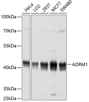
WB analysis of various sample lysates using GTX65581 ADRM1 antibody. Dilution : 1:1000 Loading : 25μg per lane


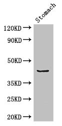
WB analysis of various sample lysates using GTX65581 ADRM1 antibody. Dilution : 1:1000 Loading : 25μg per lane
ADRM1 antibody
GTX65581
ApplicationsImmunoFluorescence, Western Blot, ImmunoCytoChemistry, ImmunoHistoChemistry, ImmunoHistoChemistry Paraffin
Product group Antibodies
ReactivityHuman
TargetADRM1
Overview
- SupplierGeneTex
- Product NameADRM1 antibody
- Delivery Days Customer9
- Application Supplier NoteWB: 1:1000 - 1:4000. ICC/IF: 1:50 - 1:200. IHC-P: 1:50 - 1:200. *Optimal dilutions/concentrations should be determined by the researcher.Not tested in other applications.
- ApplicationsImmunoFluorescence, Western Blot, ImmunoCytoChemistry, ImmunoHistoChemistry, ImmunoHistoChemistry Paraffin
- CertificationResearch Use Only
- ClonalityPolyclonal
- ConjugateUnconjugated
- Gene ID11047
- Target nameADRM1
- Target descriptionADRM1 26S proteasome ubiquitin receptor
- Target synonymsARM-1, ARM1, GP110, PSMD16, proteasomal ubiquitin receptor ADRM1, 110 kDa cell membrane glycoprotein, M(r) 110,000 surface antigen, adhesion regulating molecule 1, proteasome regulatory particle non-ATPase 13, proteasome ubiquitin receptor, rpn13 homolog
- HostRabbit
- IsotypeIgG
- Protein IDQ16186
- Protein NameProteasomal ubiquitin receptor ADRM1
- Scientific DescriptionThis gene encodes a member of the adhesion regulating molecule 1 protein family. The encoded protein is a component of the proteasome where it acts as a ubiquitin receptor and recruits the deubiquitinating enzyme, ubiquitin carboxyl-terminal hydrolase L5. Increased levels of the encoded protein are associated with increased cell adhesion, which is likely an indirect effect of this intracellular protein. Dysregulation of this gene has been implicated in carcinogenesis. Alternative splicing results in multiple transcript variants. [provided by RefSeq, Jul 2013]
- ReactivityHuman
- Storage Instruction-20°C or -80°C,2°C to 8°C
- UNSPSC41116161
Datasheet
Related products
Product group Antibodies
ADRM1 AntibodyCSB-PA001396HA01HU
ApplicationsImmunoFluorescence, Western Blot, ELISA, ImmunoHistoChemistry
ReactivityHuman, Mouse
TargetADRM1
- SizePrice
Product group Antibodies

ADRM1 AntibodyLS-C830606
ApplicationsELISA, ImmunoHistoChemistry
ReactivityHuman, Mouse, Rat
TargetADRM1
- SizePrice

Product group Antibodies
Anti-ADRM1 AntibodyHPA042266
ApplicationsWestern Blot, ImmunoCytoChemistry, ImmunoHistoChemistry
ReactivityHuman
TargetADRM1
- SizePrice

Product group Antibodies
Anti-ADRM1/ARM-1 Antibody Picoband(r)A04010-2-CARRIER-FREE
ApplicationsFlow Cytometry, ImmunoFluorescence, Western Blot, ELISA, ImmunoCytoChemistry, ImmunoHistoChemistry
ReactivityHuman, Mouse, Rat
TargetADRM1
- SizePrice
Product group Antibodies
ADRM1 Polyclonal AntibodyCAC14577
ApplicationsImmunoFluorescence, Western Blot, ELISA, ImmunoHistoChemistry
ReactivityMouse
TargetADRM1
- SizePrice
Product group Antibodies
ADRM1 antibody [AT34C2]GTX57566
ApplicationsFlow Cytometry, ImmunoFluorescence, Western Blot, ImmunoCytoChemistry
ReactivityHuman
TargetADRM1
- SizePrice
Product group Antibodies
ADRM1 antibodyGTX17239
ApplicationsWestern Blot, ELISA, ImmunoHistoChemistry, ImmunoHistoChemistry Paraffin
ReactivityHuman, Mouse, Rat
TargetADRM1
- SizePrice
Product group Antibodies
Anti-ADRM1 Antibody144-04481
ApplicationsWestern Blot
ReactivityHuman, Mouse, Rat
TargetADRM1
- SizePrice