

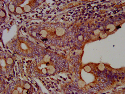
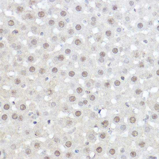
IHC image of CSB-PA600257LA01HU diluted at 1:500 and staining in paraffin-embedded human small intestine tissue performed on a Leica BondTM system. After dewaxing and hydration, antigen retrieval was mediated by high pressure in a citrate buffer (pH 6.0). Section was blocked with 10% normal goat serum 30min at RT. Then primary antibody (1% BSA) was incubated at 4°C overnight. The primary is detected by a biotinylated secondary antibody and visualized using an HRP conjugated SP system.
. Section was blocked with 10% normal goat serum 30min at RT. Then primary antibody (1% BSA) was incubated at 4°C overnight. The primary is detected by a biotinylated secondary antibody and visualized using an HRP conjugated SP system.")
.")
IHC image of CSB-PA600257LA01HU diluted at 1:500 and staining in paraffin-embedded human small intestine tissue performed on a Leica BondTM system. After dewaxing and hydration, antigen retrieval was mediated by high pressure in a citrate buffer (pH 6.0). Section was blocked with 10% normal goat serum 30min at RT. Then primary antibody (1% BSA) was incubated at 4°C overnight. The primary is detected by a biotinylated secondary antibody and visualized using an HRP conjugated SP system.
AHNAK Antibody
CSB-PA600257LA01HU
ApplicationsImmunoFluorescence, ELISA, ImmunoHistoChemistry
Product group Antibodies
ReactivityHuman
TargetAHNAK
Overview
- SupplierCusabio
- Product NameAHNAK Antibody
- Delivery Days Customer20
- ApplicationsImmunoFluorescence, ELISA, ImmunoHistoChemistry
- CertificationResearch Use Only
- ClonalityPolyclonal
- ConjugateUnconjugated
- Gene ID79026
- Target nameAHNAK
- Target descriptionAHNAK nucleoprotein
- Target synonymsAHNAK1, AHNAKRS, PM227, neuroblast differentiation-associated protein AHNAK, AHNAK-related, desmoyokin
- HostRabbit
- IsotypeIgG
- Protein IDQ09666
- Protein NameNeuroblast differentiation-associated protein AHNAK
- Scientific DescriptionMay be required for neuronal cell differentiation.
- ReactivityHuman
- Storage Instruction-20°C or -80°C
- UNSPSC41116161
Related products
Product group Antibodies
Anti-AHNAK AntibodyA307240
ApplicationsImmunoFluorescence, ImmunoCytoChemistry, ImmunoHistoChemistry
ReactivityHuman, Mouse, Rat
- SizePrice
Product group Antibodies

AHNAK AntibodyLS-C830883
ApplicationsELISA, ImmunoHistoChemistry
ReactivityHuman
TargetAHNAK
- SizePrice
Product group Antibodies
AHNAK Polyclonal AntibodyBS-13624R
ApplicationsImmunoFluorescence, ELISA, ImmunoCytoChemistry, ImmunoHistoChemistry, ImmunoHistoChemistry Frozen, ImmunoHistoChemistry Paraffin
ReactivityBovine, Canine, Human, Mouse, Porcine, Rat, Sheep
- SizePrice
Product group Antibodies
ApplicationsImmunoPrecipitation, Western Blot, ImmunoCytoChemistry, ImmunoHistoChemistry
TargetAHNAK
- SizePrice

Product group Antibodies
ApplicationsImmunoPrecipitation, Western Blot, ImmunoCytoChemistry, ImmunoHistoChemistry, ImmunoHistoChemistry Frozen
ReactivityHuman, Mouse
TargetAHNAK
- SizePrice
![ICC/IF analysis of human primary fibroblasts using GTX80164 AHNAK antibody [EM-09]. Green : Primary antibody Red : Actin Blue : DAPI](https://www.genetex.com/upload/website/prouct_img/normal/GTX80164/GTX80164_20191028_ICC-IF_2_w_23061322_864.webp)
Product group Antibodies
References
AHNAK antibody [EM-09]GTX80164
ApplicationsImmunoFluorescence, ImmunoPrecipitation, Western Blot, ImmunoCytoChemistry, ImmunoHistoChemistry, ImmunoHistoChemistry Frozen
ReactivityHuman, Mouse
TargetAHNAK
- SizePrice

Product group Antibodies
Anti-AHNAK AntibodyHPA019010
ApplicationsImmunoCytoChemistry, ImmunoHistoChemistry
ReactivityHuman
TargetAHNAK
- SizePrice