

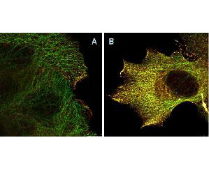
Immunofluorescence Microscopy of Mouse Anti-AKTpS473 antibody using STED nanoscopy to evaluate AKT activation and migration. Tissue: A431 cells. Antigen retrieval: Panel A: serum starved,unstimulated cells. Panel B: serum starved, EGF stimulated for 15 mins.
A massive increase in AKT-pS473 activation, as measured by intensity signal, peaked at 15 minutes and was associated with depolymerized tubulin.
Staining: Panel A shows STED data (AKT-pS473, red channel) collected simultaneously with confocal signal (a-tubulin, green channel). Upon stimulation of cells with EGF, a rapid activation of AKT is observed (Panel B) along with a coincident change in the tubulin organization (yellow signal), as well as an extensive cell shape-change (cell membrane folding) and accumulation of AKTpS473 at the cell periphery.
AKT1 Mouse Monoclonal Antibody [Clone ID: 17F6.B11]
TA396764
ApplicationsFlow Cytometry, ImmunoFluorescence, Western Blot, ELISA, ImmunoHistoChemistry
Product group Antibodies
ReactivityHuman, Mouse
TargetAKT1
Overview
- SupplierOriGene
- Product NameAKT1 Mouse Monoclonal Antibody [Clone ID: 17F6.B11]
- Delivery Days Customer14
- ApplicationsFlow Cytometry, ImmunoFluorescence, Western Blot, ELISA, ImmunoHistoChemistry
- CertificationResearch Use Only
- ClonalityMonoclonal
- Clone ID17F6.B11
- Gene ID207
- Target nameAKT1
- Target descriptionAKT serine/threonine kinase 1
- Target synonymsAKT, PKB, PKB-ALPHA, PRKBA, RAC, RAC-ALPHA, RAC-alpha serine/threonine-protein kinase, AKT1m, PKB alpha, RAC-PK-alpha, protein kinase B alpha, proto-oncogene c-Akt, rac protein kinase alpha, serine-threonine protein kinase, v-akt murine thymoma viral oncogene homolog 1, v-akt murine thymoma viral oncogene-like protein 1
- HostMouse
- IsotypeIgG1
- Protein IDP31749
- Protein NameRAC-alpha serine/threonine-protein kinase
- Scientific DescriptionAKT phospho S473 Antibody
- ReactivityHuman, Mouse
- Storage Instruction-20°C,2°C to 8°C
- UNSPSC12352203
MSDS
Related products
Product group Antibodies
AKT1/AKT2/AKT3 AntibodyCSB-PA000848
ApplicationsWestern Blot, ELISA, ImmunoHistoChemistry
ReactivityHuman, Mouse, Rat
TargetAKT1
- SizePrice

Product group Antibodies
Anti-AKT1,2,3 Antibody Picoband(r)A00024-2-CARRIER-FREE
ApplicationsFlow Cytometry, ImmunoFluorescence, Western Blot, ELISA, ImmunoCytoChemistry
ReactivityHuman, Mouse, Rat
TargetAKT1
- SizePrice
Product group Antibodies
Anti-AKT1 Antibody144-11016
ApplicationsImmunoFluorescence, Western Blot, ImmunoHistoChemistry
ReactivityHuman, Mouse, Rat
TargetAKT1
- SizePrice

Product group Antibodies
Anti-AKT1 AntibodyAMAB90834
ApplicationsWestern Blot, ImmunoCytoChemistry
ReactivityHuman
TargetAKT1
- SizePrice
Product group Antibodies

Anti-pAKT [AbAb-pAKT]AB04102-10.0
ApplicationsImmunoPrecipitation, Western Blot, ELISA
ReactivityHuman, Mouse
TargetAKT1
- SizePrice
Product group Antibodies
Anti-AKT1 AntibodyA285951
ApplicationsWestern Blot, ELISA
ReactivityHuman, Mouse
- SizePrice
Product group Antibodies

AKT1 AntibodyLS-C831478
ApplicationsImmunoHistoChemistry
ReactivityHuman
TargetAKT1
- SizePrice
Product group Antibodies

Goat anti-AKT1EB06875
ApplicationsWestern Blot, ELISA
ReactivityBovine, Human, Mouse
TargetAKT1
- SizePrice
![Akt1 antibody immunoprecipitates Akt1 protein in IP experiments. IP samples: 30 μg whole cell extract of Akt1-transfected 293T cells. A. 30 μg whole cell extract of Akt1-protein expressing 293T cell B. Control with 3 μg of preimmune Rabbit IgG C. Immunoprecipitation of Akt1 protein by 3 μg Akt1 antibody (GTX110613) 10 % SDS-PAGE The immunoprecipitated Akt1 protein was detected by Akt1 antibody (GTX110613) diluted at 1:5000. [EasyBlot anti-rabbit IgG (GTX221666-01) was used as a secondary reagent]](https://www.genetex.com/upload/website/prouct_img/normal/GTX110613/GTX110613_40051_IP_w_23060500_283.webp)
Product group Antibodies
AKT1 antibodyGTX110613
ApplicationsImmunoPrecipitation, Western Blot, ImmunoHistoChemistry, ImmunoHistoChemistry Paraffin
ReactivityHuman, Mouse
TargetAKT1
- SizePrice